Q & A: Spinous Process Fracture

Q.  My son was recently injured in a weight room accident that resulted in a C7 spinous process fracture. The neurosurgeon says not to worry about it and that he can return to sport in six weeks.  He is a very talented athlete, and he’s trying to get a college football scholarship.  I’m concerned for his safety and scholarship prospects.  Do you have any advice? -Jennifer

A.  I am so sorry to hear about your son’s injury, Jennifer. Weight lifting is such a critical component to any training program as well as for those who are trying to maintain muscle mass and general health. Sadly, accidents happen and can be very serious. However, weight training will be an ongoing critical training component if your son is to compete in football at a collegiate level.

Thank you for letting me share his imaging pictures. Viewing the imaging allows for a better discussion, but please note that I have not evaluated your son.  The following advice is only for the purpose of general discussion.

Spine_Collage

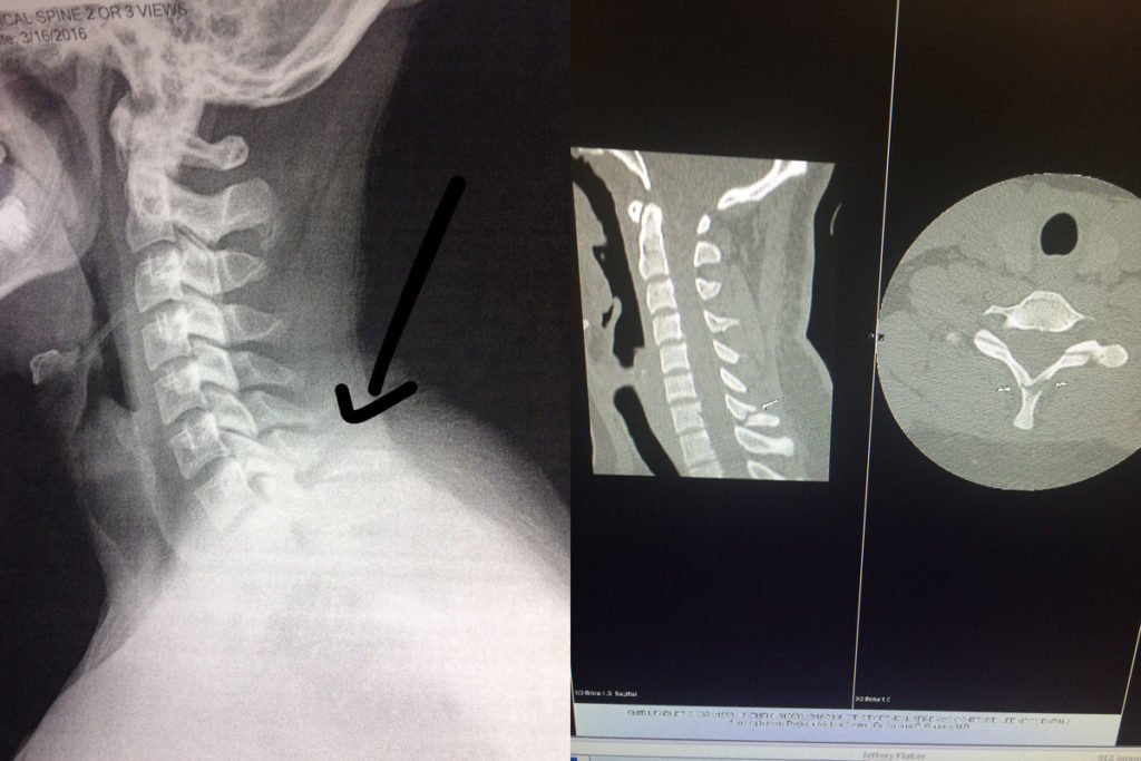
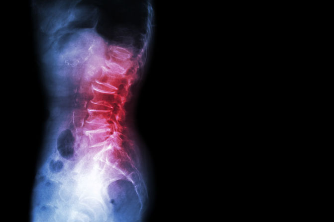
(Left: X-ray of C7 fracture. Right: CT Scan of C7 fracture.)

As you can notice on the imaging provided, only the spinous process was injured. Although close to the lamina of the vertebra (which has a primary role of protecting the spinal cord), it was not injured.  This was confirmed by the physician’s physical exam, X-rays, and CT Scan. I’m glad that the injury was taken seriously with appropriate medical follow up.  One should never take unwarranted risks when evaluating spinal injuries.

This particular fracture is also known as a Clay Shoveler’s Fracture. This occurs when the end of the spinous process is either broken off by a physical impact (as in the case of Jennifer’s son when the barbell hit his neck) OR as a result of the muscle pulling so hard that it breaks the bone and literally tears off part of the spinous process.  The role of the spinous process, particularly in the cervical region, is to help limit cervical hyper extension.  More importantly, the spinous processes are there as an attachment point for muscles and ligaments.

C7 is the largest cervical vertebrae in the neck. The C7 vertebra is right in the middle of transition from the cervical spine to the thoracic spine and is home to many fascial attachments and muscular attachments from the neck and shoulder girdle.  Many of the deeper neck extensors and rotators such as the splenius capitus and splenius cervicis as well as the trapezius, rhomboids and serratus posterior muscles have C7 attachment points.

Typical Symptoms

In most cases, there is pain immediately after the injury which is often described as a burning or “knife-like” stabbing pain.  Other symptoms include muscle tightness and sharp pain that increases with repeated activity and movement of the neck or shoulder girdle. The pain is often described as feeling similar to a severe muscle strain in the upper back.  The area of fracture as well as the nearby spinous processes is typically very tender as are the nearby muscles.

If only the spinous process is injured, you should not see any neurologic symptoms. You will likely see loss of cervical motion and possibly shoulder motion as those motions will cause muscles to pull directly on the site of injury which limits one’s desire to move.  Symptoms are typically worse with the head down or when the arms are active in front of the body (such as driving or working at the computer).

Treatment Considerations

Bone healing is a complex process and will differ significantly among individuals. Factors affecting bone healing include:  the type of fracture and the patient’s age; underlying medical conditions; and nutritional status.  For a person with good health status, the bone will take on average six to eight weeks to heal to a significant degree.  In general, children’s bones heal faster than those of adults.

However, with a spinous process fracture that is separated to this degree, the bone will most likely never heal. The bone will not re-attach itself back to vertebrae.  When this occurs, it’s known as a non-union.  Scar tissue and fascia will surround the injury site and in most cases, form a solid fibrous pseudo-union between the two pieces of bone.  In most cases, there will be no deleterious effects from the injury and healing process.  Like bone healing, this process typically takes six to eight weeks for most individuals.

Acutely there is likely to be pain, but long-term pain typically subsides. However, as with all injuries, there is the risk of chronic pain or long-term irritation to the area associated with the muscle and tendon that insert/attach onto the spinous process involved in the injury.

Pain Management

Initially, I don’t recommend treating with NSAIDS (non-steroidal anti-inflammatory medication) as there is research that indicates that the healing response could be delayed. Instead and whenever possible, I would recommend other alternatives for pain management like applying heat or ice. Typically no more than 20 minutes per hour would be recommended, and don’t apply heat or ice directly to the skin (use a towel as a barrier).

Pain medication, such as acetaminophen, is always an option if recommended by your physician. You may also want to consider utilizing topical agents, which can help to decrease pain and muscle spasms. The method of action varies greatly according to the product used.  You may find that one product works better than another.  Some of my favorite products in my medicine cabinet include:  Biofreeze Pain Relieving Gel; Arnica Rub (Arnica Montana, an herbal rub); and topical magnesium.

Another option is oral magnesium. You can take Mag Glycinate in pill form or by eating foods higher in magnesium such as spinach, artichokes, and dates. Taking additional magnesium (particularly at night) can help to reduce muscle cramps and spasming.  It is also very helpful in reducing overall muscle soreness and aiding in a better night’s rest.  Most people are deficient in the amount of magnesium they consume on a regular basis.  I recommend beginning with a dose of 200 mg (before bedtime) and increasing the dose as needed.  I would caution you that taking too much magnesium can lead to diarrhea.  Mag Glycinate in its oral form is the most highly absorbable.  Although not as absorbable, Thorne Research Magnesium Citrate and magnesium oxide can also be beneficial.

Soft tissue massage of the adjacent muscles as well as electrical stimulation can both be used to reduce pain.  If pain persists, please discuss the options with your physician.

Activity Modification and Exercise Considerations

Activity involving the head, neck, and shoulders should be limited initially. In some cases, the physician may ask that a cervical collar be worn for several weeks.  Opinions on this vary greatly and will be dependent on the exact location of the fracture.

The basic idea is to avoid heavy use of the muscles that would specifically pull on the injury site. This would include muscles that flex, extend, rotate or side bend the head as well any arm movements that move the shoulder.  This would obviously include any type of heavy lifting or placing anything on top of the injury site like a barbell.

Although this seems very limiting, you can still move the neck and shoulders. Gently perform range of motion (ROM) of the neck and shoulders to maintain full mobility. This will also help to limit pain and muscle spasms of the surrounding tissues.  The idea is to limit extreme movement, limit the movement under load, and limit the speed of movement.

I would specifically limit most activity for at least six to eight weeks to insure an adequate amount of scarring has occurred. In addition to the above noted limitations, I also wouldn’t perform any activities that would be jarring to the body (such as jogging).  If you want to maintain your cardiovascular endurance, you could peddle on a stationary bike while keeping your upper body mostly relaxed.

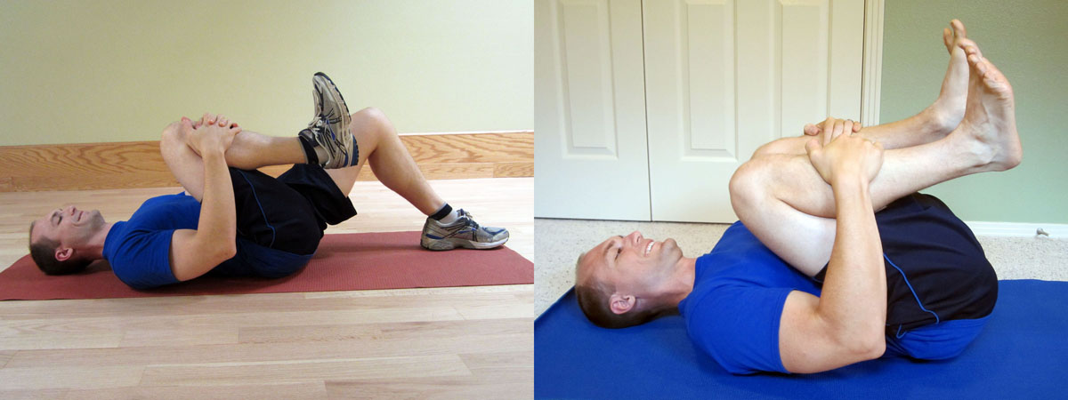
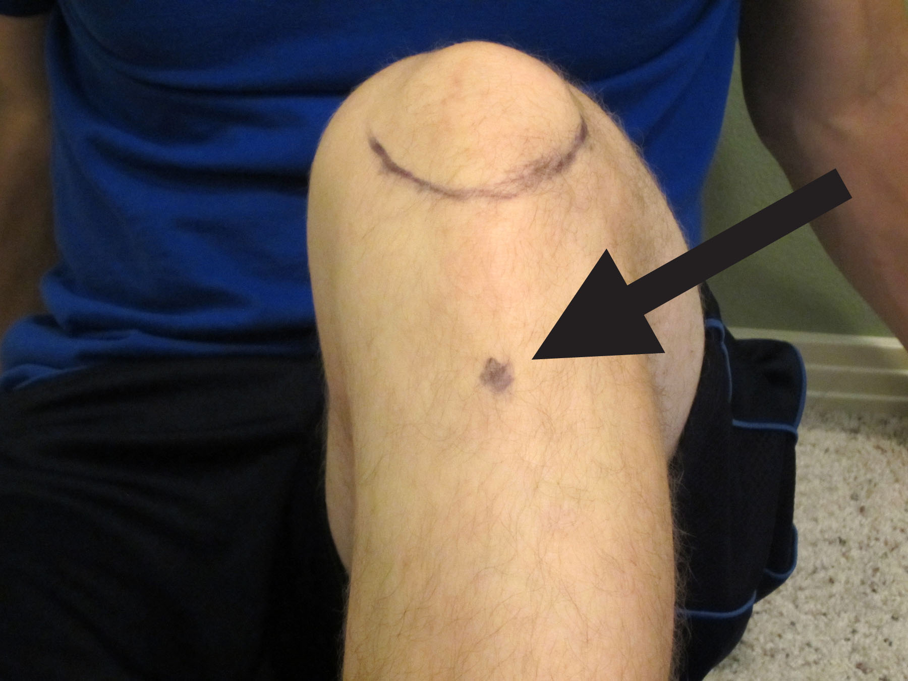
At around the four to six week mark, I would slowly start working on the cervical extensor and shoulder girdle muscle exercises (as demonstrated below).  You will want to engage the muscles symmetrically as to not cause asymmetrical pulling over the injury site.  These exercises will help to engage the muscles (the cervical extensors and scapular muscles, including the rhomboids and trapezius muscle) specifically used to stabilize the injury site.

ITYExerciseCollage

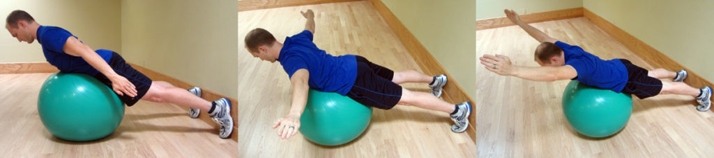
Begin by performing these I’s, T’s, and Y’s exercises on a Thera-Band Exercise Ball. Start slowly without resistance.  Keep your chin tucked and head aligned with the body.  Move your arms slowly up and down in each position of I, T, and Y.  Start with 10 repetitions for 3 second holds, and then progress the number of repetitions as long as there is no pain.

Once you can easily and pain free perform 20 repetitions with 3 second holds, add a 1-2 pound weight in each hand and start the progression initially at 10 repetitions.  Be sure to always let pain guide the progression.  The exercise should remain pain free.

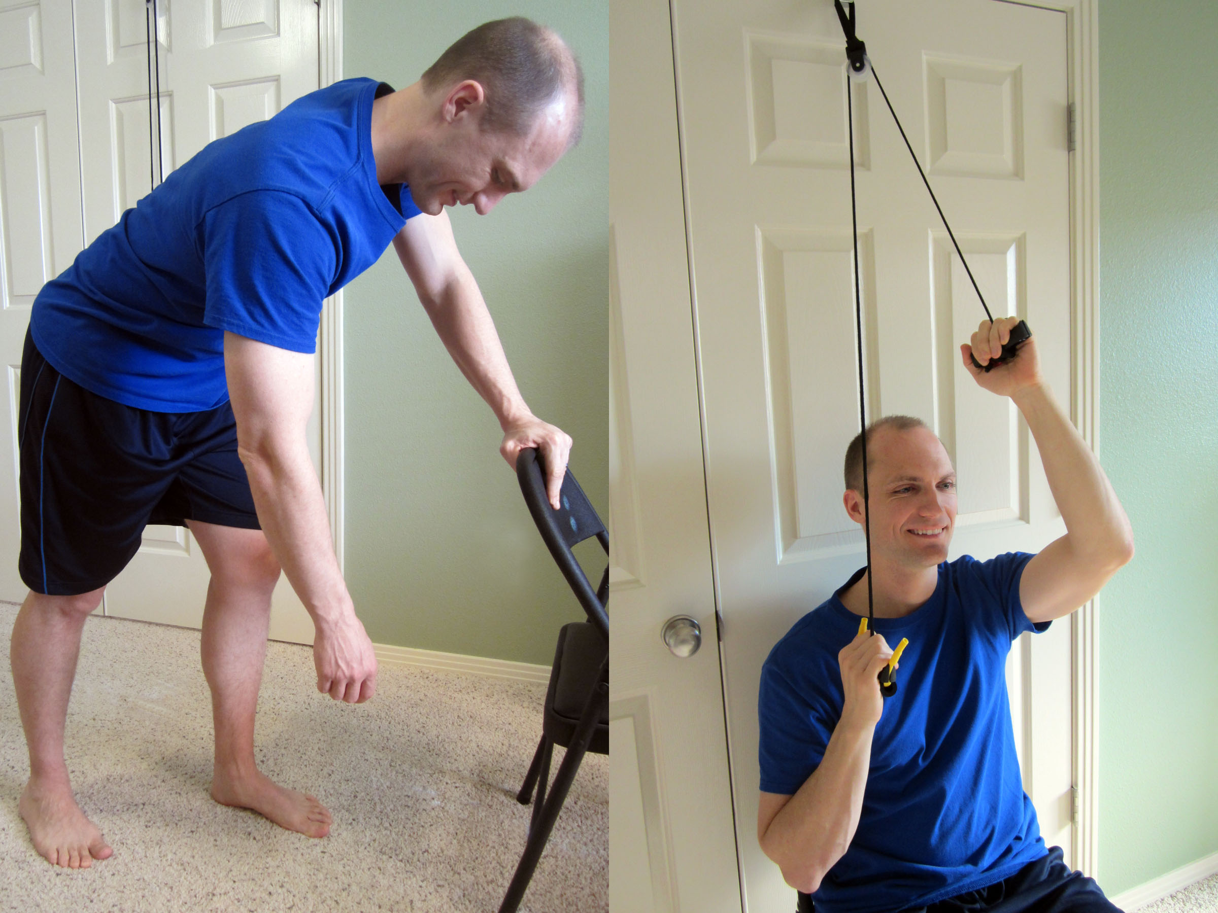
After the six to eight week mark, initiate a slow, but steady return to exercise. Start with lower extremity exercises such as the leg press, body weight lunges, squats or step ups.  During this time, progress with targeted exercises designed to strengthen the muscles of the mid trapezius, rhomboids, and neck extensors.

Let pain guide your progression. If you perform an activity that causes pain near the injury site, then that activity should continue to be avoided in the short-term.  Continue to perform exercises bilaterally to be sure that the force/pull over the injury site is symmetrical.  For example, when you start on rowing, then it should be a two-handed row and not a one-handed version.

Progression of exercise of the associated muscles should be slow and again, pain should be carefully monitored. Any pain provoking movements early in the rehabilitation phase should be avoided.  When returning back to weight training, start with a weight approximately 50% of your prior max.  Work within that weight for a week or two, and then slowly progress back to the prior weight used (depending on your symptoms).

You may also return to light jogging. If there is no increase in your pain level, then slowly progress back into full running and sprinting.

Exercises to avoid for at least twelve weeks include: barbell squats; overhead press; power cleans; squat cleans; and snatches.  Avoid anything that would put direct pressure over the injury site or exercises that include a speed and power component under load (such as the power clean).

Rehabilitation Recap

Due to the amount of information presented, I want to re-iterate the important parts of the rehabilitation process. Be sure that you have your physician’s clearance prior to resuming activity.

0-4 weeks: This is the time for pain management and activity modification. You want the area to scar down.  Exercise and activity should be limited although you should maintain full range of motion (ROM) of the neck and shoulder girdle if possible.

4-6 weeks: Initiate a cervical stabilization program and scapulothoracic exercises starting without any resistance.  Perform the I’s, T’s, and Y’s exercises on an exercise ball (as demonstrated above).  Progress with these exercises and continue with cardiovascular exercise such as riding a stationary bike.  You may also start bench pressing, but begin light and monitor your pain level.

6-8 weeks: Progress with shoulder and upper back exercises. Be sure to use two-handed movements.  Depending on your pain level and strength, lower body exercises can be initiated.  Begin with body weight exercises, and then progress to resistive exercises.  Continue to limit direct pressure over the injury site or asymmetric forces.  Lower body exercises, such as the leg press and dead lift, may be started.  Exercise should be mostly pain free.  If you experience pain during an exercise or movement, discontinue for now and then retry it in a week or two.

8-12 weeks: During this phase, slowly start tapering up on all activity.  During this time, you can progress your jogging to eventually sprinting as well as road bicycling.  You can progress back into lower extremity exercises as tolerated as long as there isn’t any significant associated pain at the injury site.  Focus on neck and upper extremities exercises with two-hands/arms bilateral exercises.  Focus on exercises that develop strength in the muscles surrounding the injury site without causing increased pain or asymmetrical force.  The preference is for two-arm movements for now.  This is also the time to start working on thoracic mobility to insure the entire vertebral chain can move freely.  Be sure to have full shoulder and cervical mobility.  Continue to avoid any specific pressure on the injury site.  If you haven’t already, you could start the following:  dead lifting; front squat; and lunges with load.

12+ weeks: Return to full activity. However, let pain and common sense continue to be your primary guide.  At this stage, you can return to light squatting.  I recommend a low-bar position as this typically lines up just below the spine of the scapula and should be below the C7 injury site.  Start single arm movements, such as a one-armed row, as well as power movements, such as the clean and snatch.  You can also initiate overhead lifting.  The key is to progress slowly and see how your body responds.  Begin with 50% of the load used prior to your injury.

If you don’t already own a copy, I would highly recommend that you purchase Starting Strength: Basic Barbell Training by Mark Rippetoe. This text should be regarded as the authority on strength training and should be used as a guide for beginners to advanced weight training enthusiasts.

Once a full return to weight training activities has been accomplished, a full return to sport should also be possible. Any sports that involve high impact, such as football or mixed martial arts, should have your physician’s approval in order to insure that the fracture is stable.  In most cases, the level of pain present will be a great indication of how stable the injury site is.

If you are unsure about how to properly progress in your training and rehabilitation, I recommend that you work with a highly qualified trainer or sports medicine professional to insure that you are performing your particular exercise and sport in a manner that will keep you safe and the fracture stable. The American Physical Therapy Association (APTA) offers a wonderful resource to help find a physical therapist in your area.  In most states, you can seek physical therapy advice without a medical doctor’s referral (although it’s a good idea to hear your physician’s opinion as well).

Good luck to your son and you, Jennifer! I hope you find this information helpful.  Thankfully, this type of injury usually results in a full return to function without many long-term deleterious effects.  The key is to allow adequate healing time, and then slowly and strategically progress back into full activity.

Have you ever suffered from a spinal fracture or avulsion fracture? Have you experienced an accident while weight training?  Please share your story below.

If you have a question that you would like featured in an upcoming blog post, please email contact@thephysicaltherapyadvisor.com. For additional health and lifestyle information, join our growing community on Facebook by liking The Physical Therapy Advisor!

Disclaimer:  The Physical Therapy Advisor blog is for general informational purposes only and does not constitute the practice of medicine or other professional health care services, including the giving of medical advice. No health care provider/patient relationship is formed.  The use of information on this blog or materials linked from this blog is at your own risk.  The content of this blog is not intended to be a substitute for professional medical advice, diagnosis, or treatment.  Do not disregard, or delay in obtaining, medical advice for any medical condition you may have.  Please seek the assistance of your health care professionals for any such conditions.

Q & A: Running Injuries, Part 2

MTA_RunningInjuries_Part2

http://marathontrainingacademy.com/running-injuries2

Marathon Training Academy

February 14, 2016

Do you have a nagging stubborn injury? Recovery and injury prevention are critical and should be performed regularly so you don’t miss valuable training time.  In Part 2 of this podcast interview with Marathon Training Academy, I discuss injury prevention and treatment for the most common running injuries.  For detailed written answers and web links for more in-depth prevention and self-treatment strategies, please refer to Q & A Running Injuries Show Notes.

Fit man gripping his injured calf muscle on a sunny dayRunning injuries that crop up during marathon training are a real bummer! Here’s part two of our Q and A with physical therapy doctor Ben Shatto.

In this episode, you will hear about injury recurrence, plantar fasciitis, knee pain, ITBS, and more. My favorite quote from this episode is, “Injury is never normal.” Listen to the podcast

Disclaimer: This blog post and podcast are not meant to replace the advice of your doctor/health care provider, or speak to the condition of one particular person but rather give general advice.

Q & A: Running Injuries

MTA_RunningInjuries

http://marathontrainingacademy.com/running-injuries

Marathon Training Academy

February 11, 2016

Do you want to meet your fitness goals? Avoiding injury is critical! In this podcast interview with Marathon Training Academy, I discuss common running injuries and self-treatment strategies.

RunningInjuryOnCalfRunning injuries! In this episode we invite Dr. Ben Shatto on the podcast and fire away with injury related questions sent in by Academy members.

You will learn when to self-treat an injury versus visiting a physical therapist, how to pick a PT, and great questions and answers about glutes, hamstrings, and muscle imbalances. Lots to love!  Listen to the podcast

Disclaimer: This blog post and podcast are not meant to replace the advice of your doctor/health care provider, or speak to the condition of one particular person but rather give general advice.

Q & A: How to Manage Spondylolisthesis Pain

Q What are some tips on how to decrease pain caused by spondylolisthesis? -Lisa

A.  Excellent question, Lisa! A spondylolisthesis (spondy) is a certain kind of back injury that is associated with a pars interarticularis defect which is part of the vertebrae. It’s often called the “scotty dog” fracture because of its shape. This condition can be congenital (from birth) or happen from trauma. It’s diagnosed slightly more in males (5% of the male population versus 3% in the female population).

It is important to note if the fracture is stable or not. If the spondylolisthesis is unstable during active motion, such as bending forward or backward, the vertebrae can actually slip and move forward or backward.

Spondylolisthesis injuries are graded I, II, III, IV, and V.

  • A Grade I defect occurs when 25% of the vertebral body has slipped forward.
  • Grade II occurs when 50% of the vertebral body slips forward.
  • Grade III occurs when 75% of the vertebral body slips forward.
  • Grade IV occurs when 100% of the vertebral body slips forward.
  • Grade V occurs when the vertebral body completely falls off which causes a spondyloptosis.

In many cases, you will never know if you have this particular condition unless an X-ray is taken. For most people, this condition is completely benign and painless (particularly, in Grade I defects).

Spondylolisthesis_XRay

If you have a Grade I or II spondylolisthesis and are experiencing pain, conservative treatment (including formal physical therapy) is usually the first form of treatment. Surgical intervention may be performed as needed in the case of a Grade II spondylolisthesis. Surgical intervention is almost always necessary in cases of Grade III or higher.

Exercise and Treatment Considerations

In case of a stable Grade I and some Grade II spondylolisthesis, exercise is an important part of the treatment strategy. There are a few items of consideration. First, obtain clearance from your medical physician. Often, a series of X-rays will be taken while you are standing and standing bending either forward or backward. This can determine if the area is stable. If so, then conservative treatment can be initiated.

When determining which motions to guide your treatment, always let pain and directional preference guide your movements. A directional preference is simply a method to identifying a pattern to the pain.  Does the pain get worse when you bend over or does it improve?  What happens when you repeat this movement?  Determine how your pain responds.  If it spreads away from the spine and down into the leg, beware that you are moving in the wrong direction.  Stop that particular movement.  If the pain improves then continue with exercises in that direction.  Never do anything that worsens your pain or symptoms.

On average, I tend to have my clients be less aggressive with lumbar range of motion, especially press-ups and backward bending. Although it’s not prohibited completely in the case of a stable injury, it’s merely a precaution as some research indicates that it may have the potential to cause more pain and worsen the pars defect.

It’s also entirely possible that the cause of pain has nothing to do with the spondylolisthesis. A thorough physical therapy evaluation should help to determine the actual cause of the pain (although, sometimes it is never truly known). The American Physical Therapy Association (APTA) offers a wonderful resource to help find a physical therapist in your area. In most states, you can seek physical therapy advice without a medical doctor’s referral (although it may be a good idea to hear your physician’s opinion as well).

The focus of the treatment and exercise is on the strengthening of the inner and outer core muscles and lumbar extensors. With the only caveat being that you may need to avoid excessive loading with the spine extended. In this instance, I recommend that you work with a highly qualified trainer or sports medicine professional to insure that you are performing your particular exercise and sport in a manner that will keep you safe and the fracture stable.

It is also important to insure proper hip and pelvic mobility so that the spine is not over worked. In cases of spondylolisthesis, insuring a normal amount of hip extension in addition to proper hamstring length and hip rotation is important. If the hip cannot fully extend during walking and running, it will cause excessive lumbar extension. You may even want to focus on having less of a lumbar curve (a posterior pelvic tilt) if your tendency is to hyper extend with an anterior pelvic tilt. Be sure to work on thoracic mobility to insure the entire vertebral chain can move freely.

Exercise is the critical component to the management of this condition. I would highly advise that you consult with a local physical therapist that has a Lumbar MedX exercise machine.  This particular machine can isolate the lumbar multifidus during exercise better than any other exercise that I am aware of.

Other exercises can be utilized to activate the multifidus. These Lumbar Extensor Exercises are designed to progressively activate the multifidus muscles (with the final exercise being the most challenging). Generally improving your core strength is a critical component to the overall treatment. My only caution is to once again avoid excessive loading in hyper extension and to take your strength progression more slowly while monitoring your symptoms.  If you perform an activity that causes worsening pain, then you will need to modify or eliminate that particular activity until it can be performed pain free.

In some cases, more flexion biased stretches would be indicated. This would be determined by the directional preference.  If extension biased exercises worsened the pain and flexion biased exercises improved the pain, then initially you would proceed with flexion biased exercises to help control pain while you progress into your core and lumbar stabilization program.  Examples of flexion biased stretches would be a single knee to chest (below left) or a double knee to chest exercise (below right).  Hold these for 20-30 seconds at a time and perform 4-5 repetitions each.

Spondylolisthesis_Collage_1200pxx450px

Good luck, Lisa! I hope you find this information to be helpful and provide some relief from the pain you’re experiencing.  For more information on treatment strategies for low back pain, please refer to How to Safely Self-Treat Low Back Pain.

Do you suffer from spondylolisthesis? Please share your best tips for pain management.

If you have a question that you would like featured in an upcoming blog post, please email contact@thephysicaltherapyadvisor.com. For additional health and lifestyle information, join our growing community on Facebook by liking The Physical Therapy Advisor!

Disclaimer:  The Physical Therapy Advisor blog is for general informational purposes only and does not constitute the practice of medicine or other professional health care services, including the giving of medical advice. No health care provider/patient relationship is formed.  The use of information on this blog or materials linked from this blog is at your own risk.  The content of this blog is not intended to be a substitute for professional medical advice, diagnosis, or treatment.  Do not disregard, or delay in obtaining, medical advice for any medical condition you may have.  Please seek the assistance of your health care professionals for any such conditions.

Q & A: What to Expect after Breaking a Collarbone

Q.  I fell snowboarding last weekend, and I broke my collarbone. My doctor says that I don’t need surgery and that I will be fine in a couple of months.  I want to be more proactive than that.  What should I do? -Shawn

A.  Thanks for the question, Shawn. I’m sorry to hear about your snowboarding accident.  Fractures of the collarbone (clavicle) are actually fairly common and typically result from falling on the shoulder, the collarbone or an outstretched hand.  Collarbone injuries are also very common in toddlers, typically due to a fall out of bed or tripping when running.

The collarbone attaches the sternum to the scapula (shoulder blade). Many important structures, such as nerves and blood vessels, lie just below the clavicle. However, these vital structures are rarely injured when the clavicle breaks.  Diagnosing a clavicle fracture is typically performed via X-ray. In certain circumstances, a CT Scan may be performed for a more thorough image of the injury.

RightClavicleFracture

Symptoms of a Collarbone Fracture include:

  • Pain in the shoulder or over the collarbone.
  • Difficulty raising your arm due to pain.
  • Slumping or sagging of the shoulder, typically downward and forward.
  • A grinding sensation in the collarbone area when attempting to raise the arm.
  • A “bump” forms over the collarbone. In severe cases, the bump will be similar to a piano key sticking up and will be mobile.
  • Bruising and swelling over the collarbone area.

Surgical Intervention

Depending on the severity of the injury, surgery may or may not be indicated. In most cases, a simple fracture (where the bones are still aligned) will not require surgery.  Even in cases of minor malalignment, most people will not undergo surgery.

Surgical fixation is most common when there is a significant displacement or malalignment present. The purpose of surgical fixation is to hold the bones in place while they heal.  The physician will either utilize a metal plate and screws or small pins to hold the bones in place.  Although the surgical hardware can be removed once the bones have sufficiently healed, often it will remain throughout a person’s lifetime.

Rehabilitation post-surgery is fairly straight forward and is similar to the following nonsurgical treatment recommendations. Please follow your physician’s recommendations after surgery as each procedure can be different.  Variations in recovery and rehabilitation can occur and are dependent on: the area that was fixed; the severity of the fracture; and the stability of the bone and fixation.  These factors will determine how quickly you can progress through the rehabilitation.

Nonsurgical Treatment Recommendations

Most collarbone fractures do not require surgery (particularly, if the fractured area retains its alignment). Your physician will assess the severity of the fracture.  This will determine how quickly you can progress through the stages of treatment.

A general time frame for healing (with an initial return to function) is on average 6 weeks for adults and about 4 weeks for children. Complete bone healing, remodeling, and a full return to all activities will take longer.  This will depend on your age, health status, and the severity of the injury.

PRICE (Protect, Rest, Ice, Compression, and Elevation)

  • Protect. In many cases, you will be advised to wear a sling (particularly, during your daily activities). This will help to protect the area from too much motion and activity while allowing the bone to heal in its proper place. It will also indicate to others that you have an injury, so that they may avoid knocking into you.
  • Rest. Allow the arm and shoulder time to rest and recover. Do not utilize the arm for activities that cause pain. Avoid excessive motions and use. As the pain subsides, you can slowly taper up the use of the arm by starting with simple daily activities.
  • Ice. Apply ice to the painful area–typically the sooner, the better. The rule for icing is to apply ice no more than twenty minutes per hour. Do not place the ice directly against the skin, especially if you are using a gel pack style. A bag of frozen peas can be ideal. Individuals with poor circulation or impaired sensation should take particular care when icing.
  • Compression. It’s not easy to apply compression in this area. In most cases, it’s not necessary. In some cases, a simple ACE wrap can be used around the shoulder and collarbone area to help reduce the pain. The wrap is typically applied to help hold the shoulder in a backward and slightly downward direction.
  • Elevation. Elevation is typically not necessary unless you are experiencing excessive swelling in the affected arm and hand. You can position the arm in a slightly elevated position by using pillows while lying on your back or on the non-affected side. This would be an excellent time to apply ice, too.

Sleeping

When sleeping, try not to lie on the affected side. Hug a small pillow for comfort.  This also promotes optimal blood flow to the shoulder area.

Pain Management

Initially begin with PRICE (Protect, Rest, Ice, Compression, and Elevation).  Pain medication, such as acetaminophen, is always an option if recommended by your physician.  You may also want to consider utilizing topical agents, which can help to decrease pain and muscle spasms. The method of action varies greatly according to the product used. You may find that one product works better than another. Some of my favorite products in my medicine cabinet include: Biofreeze Pain Relieving Gel; Arnica Rub (Arnica Montana, an herbal rub); and topical magnesium.

Another option is oral magnesium. You can take Mag Glycinate in pill form or by eating foods higher in magnesium such as spinach, artichokes, and dates. Taking additional magnesium (particularly at night) can help to reduce muscle cramps and spasming. It is also very helpful in reducing overall muscle soreness and aiding in a better night’s rest. Most people are deficient in the amount of magnesium they consume on a regular basis. I recommend beginning with a dose of 200 mg (before bedtime) and increasing the dose as needed. I would caution you that taking too much magnesium can lead to diarrhea. Mag Glycinate in its oral form is the most highly absorbable. Although not as absorbable, Thorne Research Magnesium Citrate and magnesium oxide can also be beneficial.

Supplements

Mt. Capra, an organic goat farm in eastern Washington State, offers superior quality products primarily utilizing goat based products. CapraFlex by Mt. Capra is the best bone and joint supplement I have found. It is a blend of natural herbs and spices along with glucosamine and chondroitin.  The herbal and spice formulation is designed to naturally decrease inflammation and support healing.  I recommend it to anyone recovering from an injury or attempting to prevent injury when performing at a very high level.  I personally use it, and in my practice, it has helped clients recover faster and prevent injury.  It can interfere with some blood thinning medication, so if you are on this type of medication, please check with your physician.

Vitamin D3, such as Viva Labs Vitamin D3, is critical to the absorption of calcium through the intestinal wall which is important for bone health.  Although calcium is a critical component of bone health, I cannot recommend extra supplementation because of the potential cardiac risks to over supplementation.  A healthy varied diet will typically supply adequate calcium levels (assuming that adequate Vitamin D3 levels are present for absorption and that you are avoiding drinking soda).  Vitamin D3 is also a critical nutrient in maintaining a healthy immune system.

Be sure to maintain a generally healthy diet. Give your body the needed nutrients to heal and recover quickly.

Range of Motion

Depending on the severity of the fracture, you will likely have to limit the range of motion (ROM) of the shoulder to less than 70-80 degrees of flexion (forward) and abduction (sideways) motion. You will also have to limit reaching behind your back. A person may typically wear a sling for 2-4 weeks while limiting the motion.

The initial treatment to maintain ROM is to perform the pendulum exercise (as demonstrated in the picture on the left down below). Bend forward at the waist, and let your injured arm hang down toward the ground. Make small circles with your hand, and let the momentum move your arm around effortlessly.  Try to make both clockwise and counterclockwise circles. For more instruction, please watch How to Properly Perform Pendulum Exercises.

PendulumExercise_ShoulderPulleyExercise

Around the 2-4 weeks mark, you can begin to work on regaining full forward flexion by using an over-the-door shoulder pulley (as demonstrated in the picture on the above right). Work slowly on motion for 5-10 minutes at a time. You may push though minor discomfort, but you should never experience significant pain during this process. For more instruction, please watch How to Use Shoulder Pulleys to Regain Shoulder Motion.

As flexion improves (typically after the 4 weeks mark), then you can begin to work on all other motions of the shoulder. This includes motion out to your side (abduction) and behind your back (internal rotation). The shoulder pulley can be utilized to regain these motions as well. Otherwise, you could walk your fingers up the wall when facing forward to regain flexion and sideways to regain abduction. You could use a towel to regain the motion behind your back.

Strengthening

Initially avoid lifting anything over 5 pounds. Typically around the 4-6 weeks mark (depending on severity of injury), you can begin a gentle rotator cuff strengthening program. These exercises should always be performed pain free and initially, the resistance will be very light. For specific exercises, please refer to Rotator Cuff Exercises.

The rotator cuff is a critical component to shoulder mobility. It is made up of four different muscles whose job is to make sure that the ball of the humerus (arm bone) rotates and slides properly in the socket, which is made up of the scapula. The rotator cuff allows the other major muscles of the arm, such as the deltoid and Latissimus dorsi (lats), to properly move the arm.

When there is weakness or dysfunction, it will cause rubbing of the muscle tendon on the bone. This can lead to impingement or eventually fraying and tearing. Insuring proper strength in the rotator cuff after a collarbone injury is an important component in avoiding shoulder impingement syndrome.

Return to Full Activity

Around the 8-12 weeks mark, you will likely consult with your physician to have another X-ray taken.  Once you’re cleared by your physician, you can slowly taper back into more strenuous exercises and a return to full activity.

If your injury was severe or required surgery, it may be closer to the 16 weeks mark before you can return to full activity and be cleared to lift heavier weights.  I recommend that you slowly taper back into your prior activities.  If you are experiencing pain, then back off that particular activity and try it again at a later date.

Help from a Physical Therapist (PT)

Every injury is different. If you’re experiencing pain or difficulty regaining your strength and shoulder range of motion, please consult with a physical therapist that is an orthopaedic certified specialist (OCS). The American Physical Therapy Association (APTA) offers a wonderful resource to help find a physical therapist in your area. In most states, you can seek physical therapy advice without a medical doctor’s referral (although it may be a good idea to hear your physician’s opinion as well).

Good luck, Shawn! I hope you find this information to be helpful and provide some relief from the pain you’re experiencing.

Have you broken your collarbone? What was your experience like?  Please share your best tips for recovery.

If you have a question that you would like featured in an upcoming blog post, please email contact@thephysicaltherapyadvisor.com. For additional health and lifestyle information, join our growing community on Facebook by liking The Physical Therapy Advisor!

Disclaimer:  The Physical Therapy Advisor blog is for general informational purposes only and does not constitute the practice of medicine or other professional health care services, including the giving of medical advice. No health care provider/patient relationship is formed.  The use of information on this blog or materials linked from this blog is at your own risk.  The content of this blog is not intended to be a substitute for professional medical advice, diagnosis, or treatment.  Do not disregard, or delay in obtaining, medical advice for any medical condition you may have.  Please seek the assistance of your health care professionals for any such conditions.

Q & A: How to Manage COPD

Q.  Hi, Dr. Ben. I was recently diagnosed with moderate COPD.  I “knew” I have had it for a few years, but I didn’t want to admit it.  What exercises do you recommend that I perform to keep my lungs functioning at their best capacity?  I know I need to keep active, keep a healthy lifestyle, and keep going!  I gave up smoking last week when I was diagnosed.  I am not on oxygen at this time.  Inhalers are being used to manage my COPD at this time.  I hope this will help others from being stubborn, choose to give up the smokes, and “get real” because this can happen to any of us!  Thanks, Ben!  -Linda

A.  Thanks for your question, Linda! I am sorry to hear about your recent diagnosis of COPD.  It sounds like you are already making some appropriate and much needed lifestyle changes to help to manage the disease.

Chronic obstructive pulmonary disease (COPD) is a progressive lung disease (meaning it worsens over time).  COPD is currently the third leading cause of death in the United States.  It is typically diagnosed in middle aged or older adults.  Cigarette smoking is the leading cause of COPD.  Most cases of COPD are associated with those that have smoked, used to smoke or who were around heavy amounts of smoke.  Other potential causes include: exposure to chemicals that irritate the lungs (such as air pollution, chemical fumes from paint or other industrial causes); exposure to high levels of dust and dirt; and genetic factors.

Chronic Obstructive Pulmonary Disease

Symptoms of COPD:

  • Coughing (typically producing large amounts of mucus).
  • Wheezing.
  • Shortness of breath (SOB).
  • Tightness in the chest.

With COPD, there is less air flow in and out of the lungs.  This can occur for one or several of the following reasons:

  • The airways and air sacs (alveoli) lose their elasticity.
  • Walls between the air sacs are damaged and/or destroyed.
  • Ways of the air sacks become thick and/or inflamed.
  • There is excessive mucus production in the air sacks causing them to clog.

COPD is most commonly associated with two conditions, either emphysema or chronic bronchitis.  In the case of emphysema, the walls of the air sacs are damaged and/or destroyed which leads to less air sacks (alveoli) to assist in gas exchange during breathing.  In the case of chronic bronchitis, the lining of the airways is chronically irritated and inflamed.  This leads to a thickening and excessive mucus production, which blocks the airways and causes difficulty with breathing.  Generally individuals with COPD tend to have both emphysema and chronic bronchitis.

Those with COPD are especially prone to other illnesses, such as colds, flu, and pneumonia, and they are in a high risk category.  Although controversial in western medicine, I am convinced that proper supplementation of key nutrients can assist your immune system in fighting many common illnesses.

Strategies for managing COPD:

Medication.

Your physician will likely prescribe medications to help you to manage COPD.  These will typically include anti-inflammatory medications and bronchial dilator medications.  Be sure to always follow your physician’s advice when utilizing prescription medication.

Stop smoking.

This may sound obvious, but smoking cessation is critical to managing COPD.  Smoking was likely a major contributing factor to developing COPD.  It will be very difficult to manage the disease long term if you do not stop smoking.  If a person continues to smoke, lung function and prognosis for disease management will decline at a much faster rate than average.  Many programs are available that can help you to quit smoking.  Please speak with your physician to learn more about resources in your area.

Daily activity.

Daily activity is important for everyone, but especially for those with COPD.  A sedentary lifestyle will only worsen your symptoms.  Depending on the severity of your COPD, your ability to perform certain activities will vary.  For more information, please refer to How to Jumpstart a Sedentary Lifestyle and Why You Should Walk, Not Run.

As your disease progresses, it will become increasingly difficult to remain active. Activity modification will be part of your management strategy, but continue to do what you can. It is critical to keep regular activity as part of your disease management strategy.

Exercise.

It is critically important that you engage in specific, purposeful exercise.  A thorough exercise program should include components of each of the following:

  1. Weight Training
  2. Cardiovascular Exercise
  3. High Intensity Training

Each exercise program should be specifically designed for the individual.  These three forms of exercise have significant research in improving physical function (including muscular, cardiovascular, and pulmonary function).  It is extremely important that your exercise program is properly dosed and prescribed just for you.  As your disease progresses, your dosing and exercise prescription will also change.  To adequately mange the condition, you must implement a thorough exercise program.

You will likely need assistance in designing a program specifically for your needs.  First, please speak with your physician.  Then seek a qualified physical therapist that specializes in cardiovascular disorders.  The American Physical Therapy Association offers a wonderful resource to help find a physical therapist in your area. In most states, you can seek physical therapy advice without a medical doctor’s referral (although it may be a good idea to seek your physician’s opinion as well). Another option is to seek a local cardiac and pulmonary rehabilitation program in your area. These are usually associated with your local hospital.

As your disease progresses, it will become increasingly difficult to exercise, but continue to do what you can. The intensity and duration may change and you will need additional rest. It is critical to maintain some form of exercise as part of your disease management strategy. This is an exercise is medicine approach.

Rib and thoracic mobility.

Addressing rib and thoracic mobility is one way to make quick improvements in a person’s ability to breathe and generally improve endurance and lung function. Unfortunately, this is underutilized in most COPD sufferers.

Since COPD affects the lungs, restrictions in the musculature and the skeletal system that encase the lungs will only worsen a person’s breathing difficulties. The key is to help your body move as efficiently as possible. In order to achieve this, the ribs and thoracic spine along with the associated musculature must be free to move. Any restrictions in this area will only make it more difficult to breathe.

When you sign up to receive my blog posts via e-mail, you will automatically gain access to my FREE resource, My Top 8 Stretches to Eliminate Neck, Upper Back, and Shoulder Pain. These exercises address the most common rib and thoracic restrictions, so that you can maximize your rib and thoracic mobility in order to breathe easier. Detailed photos and exercise instructions will help you to get started.  I recommend that you combine these with a daily deep breathing program.

Deep breathing.

Learning a variety of breathing techniques will also be a critical component in managing COPD. Deep breathing allows the body to uptake more oxygen. It also helps to reduce stress and anxiety. The mechanical act of breathing more deeply helps your ribs and thoracic spine to remain mobile. It also helps to train all of the muscles associated with breathing–from the diaphragm and intercostal muscles to the lesser known accessory breathing muscles (sternocleidomastoid, the scalenes, serratus anterior, pectoralis major and minor, trapezius, latissimus dorsi, erector spinae, iliocostalis lumborum, quadratus lumborum, serratus posterior superior and inferior, levatores costarum, transversus thoracis, and the subclavius).

Incentive Spirometer

When performed properly, deep breathing can help you to maintain your lung capacity. An incentive spirometer should be used regularly as part of maintaining proper lung capacity and function. It can also decrease your risk of pneumonia, which is always a concern for those with COPD.

Learning multiple methods of breathing will be important as you progress in your disease management. Each method is used for a different situation. Along with deep breathing exercises, diaphragmatic and pursed lip breathing techniques will also be beneficial.

Adequate sleep.

Poor sleep is linked to the following cardiovascular risk factors:  high blood pressure; atherosclerosis; heart failure; heart attack; stroke; diabetes; and obesity.  These all have negative effects on COPD management.

Be sure to get as much sleep as you need (typically 7-9 hours at night).  Short naps are also an excellent way to get more sleep.  I recommend that you read Michael Hyatt’s 6 Strategies to Sleep Soundly, Wake Rested, and Accomplish More.

Acupuncture is also a proven method to help people sleep better and more soundly.  It can be used to treat insomnia as well.

Reduce your stress.

Stress is a normal part of life.  Poorly managed stress or excessive stress can lead to emotional, psychological, and physical problems, such as cardiovascular disease and high blood pressure.  High stress leads to more anxiety which can affect breathing patterns.  Individuals with COPD will already struggle at times with breathing, which will worsen as the disease progresses.  High stress will only make this worse.  Regular exercise, massage, yoga, acupuncture, and Tai Chi have all been proven to reduce stress.

Maintain a healthy weight.

Additional weight makes it more difficult to move around. It also creates additional pressure in the rib cage–making breathing more difficult. The heavier you are, the more difficult it will be to exercise which is a critical component of COPD management. Obesity is associated with diabetes, high blood pressure, and coronary artery disease.  These all increase your risk of developing heart disease and have a negative effect on health and COPD management.  Studies have shown that excess body weight (not including all of the other associated medical conditions) can also lead to heart failure.  Even if you are healthy otherwise, being overweight still places you at a greater risk of developing cardiovascular and pulmonary related diseases.

Stay hydrated.

Drink more water.  The older you are, the easier it is to become dehydrated.  Proper hydration is critical to keeping lung tissue healthy. The human body is primarily made of water, which is critical for all body functions. Dehydrated tissues are prone to injury as they struggle to gain needed nutrients to heal and repair. Dehydrated tissues are less flexible and tend to accumulate waste products. Water intake supports proper brain, lung, muscle, and hormone function as well as lubrication of the joints and skin appearance.

Stay hydrated by drinking water. Try to avoid beverages that contain artificial sweeteners or chemicals with names that you can’t spell or pronounce. Drinking more water not only prevents dehydration, but it also aids in preventing urinary tract infections (UTI).

Living with and managing COPD will be a different journey for everyone.  When managed correctly, individuals with COPD can live a long time.  Although there isn’t presently a cure for COPD, treatment options continue to improve every year.  The important part is to start addressing the disease early through medication management and lifestyle changes including:  smoking cessation; healthy eating and proper hydration; stress management; breathing techniques; and optimizing your exercise and activity levels in order to maintain proper rib and thoracic mobility.

Thanks for your question, Linda!  For additional information on the topic of COPD, please visit the following:  http://www.nhlbi.nih.gov/health/health-topics/topics/copd/ and http://copd.about.com/od/copdbasics/a/copdlifeexpectancy.htm.

Do you or a loved one suffer from COPD?  Please share your experience with us.  Together we can all grow and learn how to best manage this disease.

If you have a question that you would like featured in an upcoming blog post, please comment below or submit your question to contact@thePhysicalTherapyAdvisor.com.  Be sure to join our growing community on Facebook by liking The Physical Therapy Advisor!

Disclaimer:  The Physical Therapy Advisor blog is for general informational purposes only and does not constitute the practice of medicine or other professional health care services, including the giving of medical advice.  No health care provider/patient relationship is formed.  The use of information on this blog or materials linked from this blog is at your own risk.  The content of this blog is not intended to be a substitute for professional medical advice, diagnosis, or treatment.  Do not disregard, or delay in obtaining, medical advice for any medical condition you may have.  Please seek the assistance of your health care professionals for any such conditions.

Q & A: Rehabilitation for a Total Knee Replacement

Q.  I’m going in for a total knee replacement. I wondered what type of advice you could give me so that I’m better prepared.  Thanks!  -Judy

A.  Great question, Judy! A total knee replacement is often referred to as a total knee arthroplasty (TKA).  It’s really more like a resurfacing of the knee joint.  TKA is most often used as a treatment for those suffering from moderate to severe osteoarthritis in the knee.  Other common causes for the procedure include Rheumatoid arthritis and injury.  Knee replacement is more common among women and the likelihood of replacement increases with age.

The following information on total knee arthroplasty (TKA) and rehabilitation is not meant to be all inclusive, but to serve as a starting point as to what you will likely experience during rehabilitation. Each person’s rehabilitation process is likely to vary depending on his/her particular needs.

Four basic steps are performed in a Total Knee Arthroplasty (TKA):

  1. Prepare the bone. The damaged cartilage surfaces at the ends of the femur and tibia are removed along with a small amount of underlying bone.
  2. Position the metal implants. The removed cartilage and bone is replaced with metal components that recreate the surface of the joint.
  3. Resurface the patella. The undersurface of the patella (kneecap) is cut and resurfaced with a plastic button. This is not performed with every procedure unless necessary.
  4. Insert a spacer. A medical-grade plastic spacer is inserted between the metal components to create a smooth gliding surface.

Proper rehabilitation is a critical component to overall success with this procedure.  In most cases, a physical therapist and possibly an occupational therapist will be involved in the rehabilitation process.  I tend to classify the rehabilitation into the following four major stages.

Stage I Rehabilitation

This is the initial post-surgical recovery stage in the hospital with physical therapy (typically the same day).  The goal is for you (the patient) to be upright and walking with an assistive device, such as a walker, in order to initiate very basic motion in the knee.

Although same day procedures can be performed, you will typically spend two to three days in the hospital following this procedure.  With the new surgical techniques utilized, most patients will not have any weight bearing or other mobility precautions.

There are risks of complications with any medical procedure.  The biggest risk initially is falling either from the newly operated knee giving way or complications from medication (which can cause dizziness, lightheadedness or other symptoms).  Other possible unlikely complications include: loosening of the prosthetic within the bone; blood clots; and infection.

Prior to leaving the hospital, pain management is a priority.  In addition, you need to be able to walk at least to a bathroom (typically with some assistance to maintain safety).

Four typical options for discharge:

  1. Discharge home and continue rehabilitation with in-home care.
  2. Discharge home and initiate physical therapy in an outpatient clinic.
  3. Discharge to a skilled nursing facility. This is typically reserved for those who are expected to need an extended recovery.
  4. Discharge to a sub-acute rehabilitation facility. This is typically reserved for those who may need additional medical care, but who can tolerate a more intensive rehabilitation program.

Regardless of the discharge location, the initial treatment is very similar.  Focus on pain management.  Improve your ability to ambulate.  Initiate RICE (Rest, Ice, Compression, and Elevation).  Start with basic range of motion (ROM) exercises (as described in Stage II).  Also, please refer to Total Knee Replacement Rehabilitation Exercises for many of the most common exercises you may perform depending on your individual need.

Medication Management

A major component of the rehabilitation will be to insure proper pain management.  This is typically through pain medication (both narcotic based and/or acetaminophen).  It is important to maintain proper pain management because it allows you to perform necessary activities of daily living (ADLs) as you progress through your rehabilitation.  Additional medication management may be necessary if you’re taking blood thinning medications in order to decrease the risk of blood clots.

RICE 

  • Rest. Your body has just undergone a major surgery. It will require more rest initially to properly heal and recover. Although exercise and movement will be critical to your recovery, additional rest will be needed. Limit walking on any hard surfaces, such as the flooring in grocery stores, for at least the first three to four weeks in order to minimize pain and swelling.
  • Ice. Apply ice to the painful areas especially in and around the knee. The rule for icing is to apply ice no more than twenty minutes per hour. Do not place the ice directly against the skin, especially if you are using a gel style Cold Pack. A bag of frozen peas can be ideal. Individuals with poor circulation or impaired sensation should take particular care when icing. Often, your physician will prescribe a machine known as an IceMan (or something comparable) to help with cryotherapy to reduce pain and inflammation.
  • Compression helps to prevent and decrease swelling. Swelling can cause increased pain and slow the healing response, so limit it as much as possible. You may utilize a common ACE wrap or your physician may recommend wearing TED hose which helps with swelling and decreases the risk of blood clots.
  • Elevation. When resting, keep the leg as straight as possible and elevated above the level of your heart to help reduce swelling (edema). Try not to prop anything under the knee because it would cause flexion (bending). You want the knee to move straight into full extension.

Stage II Rehabilitation

Stage II typically starts on about day three to five.  It can last up to six to eight weeks depending on your recovery.  Continue to progress with pain management, edema management, gait/ambulation training, RICE, and range of motion as initiated in Stage I.

Movement 

Regaining range of motion (ROM) is one of the primary goals with TKA rehabilitation.  Knee extension (straightening) and knee flexion (bending) must be regained as quickly as possible.  There is a limited window of time that ROM must be restored.  Aim for full recovery of ROM within the first six to eight weeks (if not sooner).

SeatedKneeExtensionStretch

Regaining both full knee extension and flexion are critical to long term success.  Once you regain full ROM, you must maintain it throughout the process.  Movement helps tissues receive the needed nutrients and can help with pain management.  Movement, including frequent toe tapping and ankle motion along with knee ROM, can also help to reduce swelling and prevents blood clots.

If the patient doesn’t regain adequate motion through the rehabilitation process, I find that knee pain usually remains a long term issue along with poor mobility.  The goal for TKA ROM is usually 0-120 degrees of motion.  (Zero meaning that the knee is perfectly straight.  The 120 degrees is how far backward you can bend the knee.)  For a point of reference, touching your heel to your buttock is usually about 150 degrees of motion.  Your knee will not have the same available ROM as a healthy knee joint, but it will be functional for most tasks.

Restoring full ROM can be accomplished in many ways.  The gentlest way is to perform heel slides by lying on your back and sliding your heel toward your buttock (as demonstrated in Total Knee Replacement Rehabilitation Exercises).  A stationary bicycle is also very helpful.  These exercises should be performed within a mild to moderate amount of pain and discomfort.  Although ROM exercises can be a little painful, they don’t damage the new joint and ultimately, the ROM will help to decrease pain.

Ambulation

In most cases, your initial ambulation will require an assistive device such as a front wheeled walker (FWW), crutches or a cane for safety and pain relief. Initially, the knee motion will be limited. Pain and swelling will affect the strength of the leg. This means that the leg could have a tendency to give way or be unsteady. This should improve quickly over the first several days, but I always recommend an assistive device initially.

Son with Elderly Father

Walking with an assistive device also gives you the opportunity to walk with a normal gait pattern which will help improve your knee’s range of motion and aid in your recovery. Your physical therapist will work with you to help restore as normal and safe a gait pattern as possible. It is common to feel or even hear clicking during ambulation and movement due to the plastic and metal components. This is normal (particularly, early in your rehabilitation when the knee is swollen and ROM remains restricted).

Strengthening

Increasing your leg strength is an important part of your recovery, but it will not necessarily be an initial focus of your recovery. The primary goals for rehabilitation initially involve proper ambulation, reducing the swelling, pain relief, and range of motion for your knee.   Adequate strength throughout the lower extremity is important. The primary focus will be on quadriceps strengthening and hip abduction (gluteus medius) strengthening. These two muscle groups tend to be the most important in regaining full functional mobility after TKA.

Stage III Rehabilitation

Similar to Stage II, rehabilitation in Stage III continues to focus on range of motion (ROM).  At this point, you would typically ride a stationary bike regularly while working to maintain full ROM (0-120 degrees).  Movement helps tissues receive the needed nutrients and can help with pain management.  Movement also reduces swelling and prevents blood clots.  This stage is typically eight to twelve weeks after the operation.

The Stage III exercise program (as demonstrated in Total Knee Replacement Rehabilitation Exercises) is designed to improve strength of the entire lower extremity while still focusing on quadriceps and gluteus medius (hip abduction) strength.  Balance and gait drills are typically introduced.  The goal is to ambulate without any noticeable abnormalities from the TKA and to reduce the risk of falls (particularly if you’re no longer using an assistive device).

During Stage III rehabilitation, I would generally expect that you have proper pain management without the use of narcotic medications.  Mobility for basic daily tasks is usually quite good and the intensity of the rehabilitation has increased.  In this stage, you’re working hard on self-rehabilitation and may be continuing formal rehabilitation in an outpatient physical therapy clinic.

Stage IV Rehabilitation

Stage IV usually occurs four to nine months after the operation.  Exercises should be designed to help maximize your mobility and desired mobility outcome/goal.  Increased exercise intensity and more dynamic balance training are introduced.  A return to full functional mobility is the desired outcome.  In this stage, a majority of the exercises are performed independently.  You may intermittently consult with a physical therapist to progress in rehabilitation to insure that you’re meeting your goals.

Each person’s rehabilitation process is likely to vary depending on his/her particular needs. Always be open and honest with the medical professionals who are assisting you. If you feel as though something is not quite right, don’t hesitate in discussing the issue.

During your recovery, don’t forget the basics! Proper hydration and consuming nutrient dense food is critical to providing your body with the necessary building blocks to optimize your recovery. Proper hydration is critical in reducing your risk of blood clots.  It also helps the body to deliver much needed hydration and nutrients to your healing tissues.

Physical therapy will be an important component to your recovery and rehabilitation.  If you don’t already have a physical therapist that you are comfortable working with, The American Physical Therapy Association offers a wonderful resource to help find one in your area.  Depending on how you progress during your rehabilitation, you will likely work with multiple physical therapists while at the acute care or rehabilitation hospital, during in-home care, and/or at an outpatient clinic.

Thank you, Judy, for your question.  Good luck with your procedure!  I hope your rehabilitation for your knee goes smoothly and that you recover quickly!

Have you undergone a total knee arthroplasty (TKA)?  What was your experience like?  Please share your best tips for recovery.

If you have a question that you would like featured in an upcoming blog post, please comment below or submit your question to contact@thePhysicalTherapyAdvisor.com.  Be sure to join our growing community on Facebook by liking The Physical Therapy Advisor!

Disclaimer:  The Physical Therapy Advisor blog is for general informational purposes only and does not constitute the practice of medicine or other professional health care services, including the giving of medical advice.  No health care provider/patient relationship is formed.  The use of information on this blog or materials linked from this blog is at your own risk.  The content of this blog is not intended to be a substitute for professional medical advice, diagnosis, or treatment.  Do not disregard, or delay in obtaining, medical advice for any medical condition you may have.  Please seek the assistance of your health care professionals for any such conditions.

Q & A: My Top 5 Tips on How to Self-Treat Osgood-Schlatter Disease

Q.  For the past several months, my 12 year old son has been experiencing pain below his knee cap just to the top of his shinbone. He typically experiences pain when playing soccer or other sports.  The pain goes away when he rests.  It’s getting to the point that it’s hard for him to participate in sports.  He hasn’t had any injuries.  What do you recommend? -Chandra

A.  Thanks for your question, Chandra. I’m sorry to hear about your son’s knee pain.  Unfortunately, it’s a common problem in children (particularly, boys).  The symptoms that you are describing sound like Osgood-Schlatter disease although other potential causes could be Patellar Tendinitis or Patellar Femoral Pain Syndrome. Due to his age, gender, and activity level, it’s most likely Osgood-Schlatter disease.

Osgood-Schlatter disease (OS) is an overuse injury with pain located just below the knee where the patellar tendon ends (inserts) on the Tibial tuberosity. A boney nob may start to develop in this area and usually occurs as the condition progresses.  This occurs due to the excessive stress on the skeletal system during a period of rapid growing combined with stress from activity of the quadriceps muscle pulling on the patellar tendon at its insertion site on the bone.  If a bump forms, don’t worry as it is benign with OS.  It will likely continue to be there as your child ages.  There are typically no long term effects for this condition except for pain over the raised area of bone when kneeling on a hard surface.

OsgoodSchlatter_TibialTuberosity

Risk Factors for Osgood-Schlatter disease include:

  • Boys who are 11-13 years old.
  • Period of rapid skeletal growth.
  • Adolescents who regularly participate in running, jumping, and sports with many cutting or rapid changes in direction.

Symptoms for Osgood-Schlatter disease include:

  • Pain and possible swelling below the knee located near the top of the shinbone.
  • A bump or boney growth on the front of the upper shin at the Tibial tuberosity that is typically painful to touch.
  • Pain with running, jumping, and cutting sports.
  • Decreased pain or no pain at rest.
  • Possible loss of knee range of motion, typically flexion.
  • Possible pain and tightness with quadriceps stretching due to the pull on the patellar tendon.
  • Loss of quadriceps strength which mostly due to pain inhibiting its use.
  • Painful kneeling.

OS typically begins with mild soreness. The condition is typically self-limiting because of pain.  Continuing to push through the pain can cause significant long-lasting pain resulting in limited function.  Early intervention is usually very helpful.

My Top 5 Tips on How to Self-Treat Osgood-Schlatter Disease:

RICE

RICE, which stands for Rest, Ice, Compression, and Elevation.

Rest. In this case, rest would indicate tapering down from your regular exercise activity or any activity that involves running and jumping or that is causing knee pain.

Ice. Apply ice to the painful area–typically the sooner, the better. The rule for icing is to apply ice no more than twenty minutes per hour. Do not place the ice directly against the skin, especially if you are using a gel pack style.  A bag of frozen peas can be ideal.  Individuals with poor circulation or impaired sensation should take particular care when icing.

Compression helps to prevent and decrease swelling. Swelling can cause increased pain and slow the healing response, so limit it as much as possible.  You can utilize a common ACE wrap.  If you have a friend who is medically trained, many different taping techniques can also assist in decreasing swelling.  You may also consider a compression sleeve or garment.

Elevation. If there is swelling, then elevating the leg may be helpful.

If it hurts, don’t do it!

Modify the activity or discontinue it completely. If your knee is hurting when performing your sport or activity whether you are running, jumping or even weight lifting, then discontinue the activity temporarily.  If you are able to modify and perform the exercise or activity pain free, it would be okay to continue in most cases.

Improve your range of motion (ROM).

The primary goal of a rehabilitation program is to regain full pain free ROM for knee flexion (bending) and extension (straightening). This can be accomplished in many ways.  Perform heel slides by lying on your back and sliding your heel toward your buttocks.  Consider using a pole to assist in performing deeps squats.  Another stretch would be to hang onto a pole or a doorframe, and bring your heel toward your buttocks as you perform a quadriceps stretch.

These exercises should only be performed within a mild to moderate amount of discomfort in order to regain full pain free ROM. If you experience muscle tightness and soreness, I recommend using a foam roller to assist with any myofascial symptoms.  To learn how to use a foam roller, please refer to Foam Rolling for Rehabilitation.

Work on strengthening.

The primary goal of a strengthening program is to work on the quadriceps and glutes medius (hip abduction). Weight lifting is an appropriate choice, but you may have to initially limit your range of motion (ROM).  Most of my clients begin on a non-weight bearing program, then progress to partial weight bearing, and eventually, full weight bearing.  The more severe the symptoms, the longer it will take for an individual to progress to more difficult exercises or to increase the resistance (load).

To initiate a physical therapy program, please refer to Osgood-Schlatter Disease Rehabilitation Exercises.  This exercise guide is designed to address the muscles that I find to be the weakest in most individuals.  The exercises are listed from easiest to most challenging and are designed to primarily improve quadriceps and hip strengthening.  Initially, focus on full ROM.

Weight training exercises (with machine weights or free weights) should be geared toward general leg strengthening and may include: squats; leg press; hip abduction machine; step ups; dead lifts; and straight leg dead lifts. If further instruction is needed, search YouTube to watch the proper technique for a specific exercise.  Do not perform any seated knee extension exercises.

Kinesiological taping.

Besides pain relief, the purpose of Kinesiological tape is to provide compression over the Tibial tuberosity and to facilitate additional blood flow to the area. It also provides proprioceptive input which can help the knee during activity.  I have had luck using Kinesio Tape, Rock Tape, and Mummy Tape brands. There are many other useful taping techniques which utilize different forms of tape.  (You could also utilize Spider tape or KT TAPE.) To visually learn how to apply the tape, please refer to Kinesiological Taping for Osgood-Schlatter Disease.  For application and removal tips, please refer to Skin Care with Taping.

How to Prevent Osgood-Schlatter Disease:

Be sure to schedule rest between athletic seasons, athletic events, and higher volume training periods. Rest is a critical factor as an adolescent’s body undergoes physical changes.  OS is typically associated with overtraining/overuse in combination with a rapidly growing skeletal system.  There are times when an adolescent needs to rest.  It’s important not to encourage an adolescent to play sports through the pain without adequate recovery, rest, and if necessary, an evaluation by a qualified physical therapist or physician.

As part of a prevention and cross training strategy, implement the exercises demonstrated in the Osgood-Schlatter Disease Rehabilitation Exercises.  A proper warm up and cool down is critical when performing in an athletic event or a significant training session.

  • Warm up prior to exercise. I recommend that you increase your normal warm up time by at least 10 minutes in order to increase blood flow to the area. This allows better mobility and also promotes healing as movement is necessary to bring in the nutrients. Use a stationary bike or the rower machine initially to get the muscles warm and the knee joint more lubricated. Then work on moving into a deep squat position multiple times as part of the warm up. You may need to hang onto a beam or a pole to take some pressure off of your knee as you move in and out of the squat.
  • Cool down. After performing your exercises, take extra time to cool down and stretch. Use either a stationary bike (at a causal/slower pace) or the rower machine. Both are reduced weight bearing exercises that promote movement and circulation to the knee as well as increasing ROM. This is the perfect time to work through my recommended rehabilitation exercises.

Thank you, Chandra, for your question! I hope these tips will help you assist your son in his recovery and get him back to playing sports as quickly as he is able to without pain!  If the pain continues, please seek additional assistance from a qualified physical therapist or physician.  The American Physical Therapy Association offers a wonderful resource to help find a physical therapist in your area.

Do you know an adolescent who may be suffering from Osgood-Schlatter disease? If so, please share my recommendations on how to address this common and treatable condition.

If you have a question that you would like featured in an upcoming blog post, please comment below or submit your question to contact@thePhysicalTherapyAdvisor.com. Be sure to join our growing community on Facebook by liking The Physical Therapy Advisor!

Disclaimer:  The Physical Therapy Advisor blog is for general informational purposes only and does not constitute the practice of medicine or other professional health care services, including the giving of medical advice. No health care provider/patient relationship is formed.  The use of information on this blog or materials linked from this blog is at your own risk.  The content of this blog is not intended to be a substitute for professional medical advice, diagnosis, or treatment.  Do not disregard, or delay in obtaining, medical advice for any medical condition you may have.  Please seek the assistance of your health care professionals for any such conditions.

Q & A: How to Self-Treat Osteopenia

Q.  I had my annual visit with my physician, and she says that I have osteopenia. She recommended that I start taking calcium and maybe even another prescription to help. Isn’t there a better way to treat this without the use of drugs? -Mary

A.  Thank you, Mary, for this fantastic question! Osteopenia can be a very serious condition and is the precursor to osteoporosis. Osteopenia is defined as lower than normal bone density, but not to the severity of osteoporosis.

Osteopenia and osteoporosis significantly increase your risk of a bone fracture if you fall. When the condition is severe, the fracture can occur spontaneously from normal activities. Nearly 20% of all hip fractures in the elderly will result in death within the first year, and 50% of people will never regain their prior level of function after suffering from a hip fracture. Treating osteopenia and osteoporosis is a critical first step in reducing fractures and maintaining independence as a person ages.

Osteopenia can occur for many different reasons. Risk factors include:

  • Side effects of medications, such as chemotherapy or chronic corticosteroid use
  • Eating disorders
  • Radiation exposure
  • Gender – Females are more likely to experience bone loss.
  • Certain ethnic groups – White Anglo-Saxons are more likely to have thin bones.
  • Smoking
  • Prior family history
  • Alcoholism
  • Chronic soda consumption
  • Having a very thin build
  • Lack of physical activity

Osteopenia and osteoporosis are typically diagnosed via a bone density scan, which is recommended for woman over the age of 65. Depending on the risk factors for both men and women, your physician may order the scan at different times. For more information on a bone density scan, please refer to Q & A: Which Type of Imaging to Use?

Mary, you are correct. There are many options to help manage this condition other than prescription medications. I highly suggest that you speak with your physician about all available treatment options. The following recommendations are helpful in managing and treating osteopenia, but there may be more complicated internal issues affecting your body’s ability to improve bone density. Hormonal imbalances (such as low testosterone), menopause related hormone changes, and thyroid related issues (among others) may affect bone density. All possible hormone regulation issues should be screened for and addressed as you implement the following strategies.

Senior couple in the gym. How to Self-Treat Osteopenia.

How to Self-Treat Osteopenia:

1. Weight Training

Weight training (particularly barbell training) loads the skeleton progressively over time, which can decrease bone loss and increase bone mineral density. It also positively affects hormones, such as human growth hormone (HGH) and testosterone, needed to improve bone density and muscle strength. Barbell training is the most effective method due to the progressive load on the skeletal system, and the muscle pull being exerted on the bone, which also stimulates bone formation.

Weight training or resistance training (other than through barbell training) can also be beneficial. An example of weight or resistance training would be any type of pushing or pulling exercise that exerts a force on the muscle, which causes it to work harder than it would normally. Although any form of resistive exercise would be beneficial, the most effective exercises either activate large muscle groups and/or load the skeletal system. Examples include squats, lunges, and dead lifts.

2. Weight Bearing Activities

This refers to any activity, such as walking, jogging, and hiking, performed against gravity. Biking and swimming are examples of exercises that are not weight bearing and therefore, they aren’t as beneficial for bone development.

3. Vitamin D

Vitamin D is critical to the absorption of calcium, which is important for bone health. Although calcium is a critical component of bone health, I cannot recommend extra supplementation because of the potential cardiac risks to over supplementation. A healthy varied diet will typically supply adequate calcium levels (assuming that adequate vitamin D levels are present for absorption and that you are avoiding drinking soda).

4. Avoid Soda

Excessive soda intake (particularly diet soda) has been linked to poor bone density. The exact cause isn’t entirely clear. It’s debatable as to whether it’s a sign of an unhealthy lifestyle or that the additives, acids, and artificial sweeteners affect the bone density (but it’s likely both factors). What is clear is that too much soda is bad for your health and bad for your bones.

5. Creatine

Creatine monohydrate is helpful for increasing skeletal muscle size, strength, and power production. Increased muscle strength means increased muscle pull on the bones, which causes them to grow stronger. It may also have the added benefit of a preventative effect for dementia and neurodegenerative diseases. I recommend taking 5 g per day, and I typically consume it in 6-8 week cycles.

6. Magnesium

Magnesium is a critical component of bone health and health in general. I also use magnesium as a sleep and recovery aid. I consume it at night to help me sleep. Magnesium can also reduce muscle soreness, cramps and/or spasms. You can take Mag Glycinate in pill form or by eating foods higher in magnesium such as spinach, artichokes, and dates.

Most people are deficient in the amount of magnesium they consume on a regular basis. I recommend beginning with a dose of 200 mg (before bedtime) and increasing the dose in 100 mg intervals as needed. I would caution you that taking too much magnesium can lead to diarrhea. Mag Glycinate in its oral form is the most highly absorbable. Although not as absorbable, Thorne Research Magnesium Citrate and magnesium oxide can also be beneficial.

7. Stop Smoking

If you smoke, please stop. It not only affects your bone density, but it has negative effects on every other body system. It also increases your risk of cancer and heart disease.

8. Eat Healthy

Do not eat anything that comes in a package. Most of our food should be from low sugar fruits and vegetables as well as protein and healthy fats (primarily from plant sources such as avocados and coconut or olive oil). Any animal fat should be from organic and grass fed animals. Your body tissue needs nutrients to be able to perform at a high level.

Avoid processed food as much as possible. Limit sugary food and add more protein and healthy fat in your diet. Maintaining a diet with adequate healthy fats is essential in providing the nutrients to support all hormone function in the body as well as support the brain and nervous system. Adequate protein intake is necessary to support muscle health and development. A healthy diet equates to healthy bones.

9. Avoid Excessive Alcohol Consumption

A moderate amount of alcohol has been shown to have cardiovascular health benefits. Excessive amounts can lead to kidney and liver related issues as well as negatively affect bone density. The general recommendation is to consume no more than 1-2 servings of alcohol per day.

Mary, you should be able to affectively treat osteopenia without prescription medications by implementing these strategies, avoiding known risk factors when possible, and addressing any potential hormonal related issues. Thanks again for the question!

How has osteopenia or osteoporosis affected your life? Which strategies can you implement to improve your condition? Please leave your comments below.

If you have a question that you would like featured in an upcoming blog post, please comment below or submit your question to contact@thePhysicalTherapyAdvisor.com. Be sure to join our growing community on Facebook by liking The Physical Therapy Advisor!

Disclaimer:  The Physical Therapy Advisor blog is for general informational purposes only and does not constitute the practice of medicine or other professional health care services, including the giving of medical advice. No health care provider/patient relationship is formed. The use of information on this blog or materials linked from this blog is at your own risk. The content of this blog is not intended to be a substitute for professional medical advice, diagnosis, or treatment. Do not disregard, or delay in obtaining, medical advice for any medical condition you may have. Please seek the assistance of your health care professionals for any such conditions.

Q & A: Which Type of Imaging to Use?

Q.  Your previous post about MRI’s brought up a question I have always wondered. When do you need an MRI versus a CAT Scan versus an X-ray? How do you know which type of imaging you need? -Jeanne

A.  Thanks, Jeanne, for a great follow up question from Q & A: Do I Need an MRI for Low Back Pain? Many different types of imaging, such as an X-ray, CT Scan, and MRI, can be used depending on the situation and the structure being assessed. Let’s go through a quick synopsis of the basics on imaging. In addition, I will review the differences between a bone scan and a bone density scan, which is very important in diagnosing osteoporosis.

close up of male doctor holding x-ray or roentgen image

An X-ray, also known as radiography or a Roentgen ray, is an electromagnetic wave. The images produced show the parts of your body in different shades of black and white. This is because different tissues absorb different amounts of radiation (X-ray). The more radiation absorbed, the whiter it appears on the film. For example, calcium in bones absorbs X-rays the most, so bones appear white. Fat and other soft tissues absorb less and appear gray. Air absorbs the least, so the lungs appear black. If you have any metal implants, they will appear the most white.

X-rays are typically used to examine bones for a fracture or injury. Chest X-rays are often used to quickly spot pneumonia. Mammograms use X-rays to search for breast cancer. Fluoroscopy is a type of live action X-ray in which the results are delivered in real time. This is commonly used during certain types of spinal injections.

A computed tomography CT Scan (also known as a CAT Scan) uses X-rays to produce detailed images of the structures of the body. The CT scanner sends the X-rays through the body part/area being studied by taking very thin slices (images) of the targeted organ or area. They are then grouped back together by a computer to make a comprehensive image. CT Scans are best suited for viewing bone injuries, diagnosing lung and chest problems, and detecting cancer. CT Scans are widely used in Emergency Departments (ED’s) because the scan takes fewer than 5 minutes to perform depending on location of the scan. CT Scans produce more radiation than a typical X-ray or an MRI.

An MRI (Magnetic Resonance Imaging) is best suited for examining soft tissues, such as ligaments and tendons, spinal cord injuries, and brain tumors. Unlike an X-ray or a CT Scan, a MRI can take 30 minutes or more depending on the area and the detail of the scan. Like a CT Scan, the MRI takes images in slices, and then a computer recreates the information to give a comprehensive image. The smaller the slices taken, the more detailed and accurate the image meaning that the scan will take longer and cost more. An MRI typically costs more than a CT Scan. One advantage of an MRI is that it doesn’t use radiation like a CAT Scan.

A bone scan is a nuclear imaging test for looking at specific bone related injuries or disease. One advantage of a bone scan is that it can often discover a problem days to months earlier than a regular X-ray test.

During the scan, a radioactive substance called a “tracer” is injected into a vein in your arm. The tracer travels through your bloodstream and into your bones. Then a special camera takes images of the tracer in your bones. Areas that absorb little or no amount of tracer appear as dark or “cold” spots. This could show a lack of blood supply to the bone or certain types of cancer.

Areas of fast bone growth or repair absorb more tracer and show up as bright or “hot” spots in the images. Hot spots may point to problems such as arthritis, a tumor, a fracture or an infection. The level of detail is not always as good for a bone scan. However, bone scans can be very helpful for your physician in order to gain a proper diagnosis if you are experiencing unexplainable skeletal pain, bone infection or a bone injury that can’t be seen on a standard X-ray.

A bone density scan, also known as a Dual-energy X-ray absorptiometry (DXA, previously DEXA), is not the same as a bone scan. This particular scan measures the density of the bones by using two X-ray beams, each with different energy levels. One beam is high energy while the other is low energy. The amount of X-rays that pass through the bone is measured for each beam. This will vary depending on the thickness of the bone. Based on the difference between the two beams, the bone density can be measured.

The actual radiation exposure is low and typically less than a standard X-ray. A bone density scan is not only quick and safe, but it’s very important in diagnosing osteoporosis, which is the thinning of bones to the point they can become brittle and break. The National Osteoporosis Foundation recommends bone density scans for:

  • Women who are 65 years and older
  • Men who are 70 years and older
  • Those who have broken a bone after the age of 50
  • Women of menopausal age with risk factors for osteoporosis
  • Post-menopausal women under the age of 65 with risk factors for osteoporosis
  • Men between 50-69 years old with risk factors for osteoporosis

Thanks, Jeanne, for the question! The only way you can insure that you are receiving the best possible care for you and your loved ones is to understand more about your body and the medical options available today. Then you can research certain topics more thoroughly and have a complete, straight forward dialog with your medical providers.

I strongly believe that it is critical for all individuals to increase his/her knowledge base on basic medicine, health, fitness, and nutrition. My ultimate goal for The Physical Therapy Advisor is to help you in providing that education. What are your pains? What questions do you have? Please submit them to contact@thePhysicalTherapyAdvisor.com. I look forward to providing you with useful and practical types of “how to” information and to answer your health related questions.  You can achieve optimal health!

Don’t forget subscribe to my e-mail newsletter! I will send you weekly posts on how to maximize your health, self-treat those annoying orthopaedic injuries, and gracefully age. To thank you for subscribing, you will automatically gain access to my FREE resource, My Top 8 Stretches to Eliminate Neck, Upper Back, and Shoulder Pain.

Be sure to join our growing community on Facebook by liking The Physical Therapy Advisor where you will receive additional health and lifestyle information!