Does Kinesiological Tape Really Work?

I was first introduced to Kinesiological (Kinesio) style taping during a continuing education class in 2005.  Since then, this style of taping has exploded in the mainstream with athletes and celebrities alike wearing it.  Research on this modality was initially scant.  Today there are over 540 published studies with even more antidotal testimony!  Although the research is ongoing and published regularly, I get asked all the time about what can you use Kinesiological tape for and how exactly it works.  I will review three theories on how Kinesiological tape actually works and the different reasons for using it.

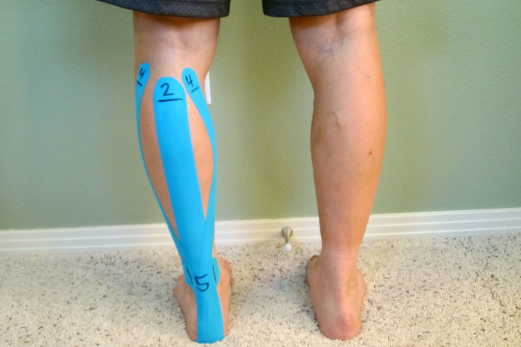
Kinesiological Taping For Achilles Tendinitis

How does Kinesiological Tape Work?

  • Sensory Theory (also known as the Gate Control Theory) – The basic premise of this theory is that the Kinesiological tape, when applied to the skin, activates sensory receptors present on the skin. These receptors relay information to/from the brain.  Since the sensory receptors are faster than other types of receptors (such as pain) the brain reacts to the sensory information first.  This can lead to altered movement patterns and awareness to the area.  It can also allow for decreased sensations of pain.  This theory is similar as to why we think topical analgesics (such as Biofreeze) work as well as why transcutaneous electrical nerve stimulation (TENS units) can reduce pain.
  • Circulatory Theory – The basic premise of this theory is that the when the Kinesiological tape is applied with little tension it forms convolutions in the skin. These convolutions create channels and reduce pressure within the tissues, lymph system, and circulatory system which aids in blood and lymphatic flow.
  • Muscle Activation Theory – The premise of this theory is that when the Kinesiological tape is applied at different levels of tension on the skin over the muscles and tissues, the Kinesiological tape can either mechanically or neurological increase or decrease muscle activation via a nervous system response.

Presently the truth behind Kinesiological taping is that we really don’t know exactly how or why it works.  Based on my clinical experience, I believe that the Kinesiological tape interacts with the body’s tissues in different ways depending on how and where the Kinesiological tape is applied.  Depending on how the Kinesiological tape is utilized, any one or combination of the three theories is likely correct.

Research states that Kinesiological style taping is at least as effective as other minimal interventions for musculoskeletal pain which may include topical analgesics like Arnica montana (Arnica Rub) or Biofreeze.

Research has also shown that taping can reduce pain acutely within first week of injury and possibly even in cases of pain which has been present for as long as three to four weeks (or longer).

Research concludes that there are some improvements in muscle and joint range of motion (ROM) and a reduction of pain.  However, because the improvement isn’t significant or long standing it shouldn’t be used as a standalone intervention.  Kinesiological taping is best used in combination with other interventions (such as manual therapy based techniques and exercise) which have been proven to affect outcomes over the long-term.

When Kinesiological tape is used in conjunction with other treatments, it may help speed up the recovery as the Kinesiological tape can allow for other techniques to be utilized more effectively due to the loss of pain, swelling or easing of movements.

Although there have been hundreds of research studies performed, the effectiveness of Kinesiological tape is still questioned primarily due to issues with research design.  Many of the taping applications are based on theory and not science.  Since there isn’t a consistent application pattern, the research studies aren’t comparing the same techniques over large enough patient populations to show a true effect.  In addition, researchers still need to quantify how much tension is actually being used and the actual Kinesiological taping technique being used versus other types of techniques or interventions.

Unfortunately, without better quality research it’s unclear if the results shown in many of the studies are due to an actual effect or through placebo.  For now, none of the studies show significant benefits regarding long-term pain alleviation or improved strength or range of motion (ROM).  So for now, the main conclusion is that Kinesiological taping should be used only as a short-term treatment in combination with other treatments.  I personally use Kinesiological tape, and I often find that my clients experience success with specific tape applications for different conditions.

What do you use Kinesiological Tape for?

  • Reduce pain
  • Reduce swelling and edema
  • Support muscles and joints
  • Reduce muscle tension
  • Increase strength
  • Improve performance
  • Enhance or correct movement patterns through stimulation of sensory receptors
  • Provide compression
  • Enhance healing by slightly lifting skin away from sore or injured tissues to improve blood flow and lymphatic drainage
  • Support injured joints and muscles without impeding range of motion (ROM)

There are dozens of companies’ now manufacturing Kinesiological style tape.  There are many high quality tapes being manufactured with all kinds of colors, designs, and variations in adhesive quality and elasticity.  Personally, my favorite brands are based on price point and actual use.  This includes ease of application and how long the Kinesiological tape actual stays on.  The brands I use most include:  Kinesio Tape, RockTape, Thera-Band Kinesiology tape, Spider tape, and KT TAPE.  Although there are many more brands available, I have not personally used them.

There are many different applications Kinesiological taping can be used for.  Like any treatment modality, it can work wonders for one person and do nothing for another, but should we really be surprised?  Many of our most popular pharmaceuticals are no different.  Kinesiological taping has the benefit of rarely having significant side effects.  There are rarely side effects to utilizing this style of tape as the products utilized tend to be very hypoallergenic.  Occasional skin irritation has been the only noted side effect I have known of in my clinical experience.  In order to avoid skin related issues (including application and removal tips), please refer to Skin Care with Taping.

I have found success with utilizing Kinesiological taping as an adjunct treatment strategy and as part of a complete treatment approach.  If you are interested in specific applications for different conditions, please see the following in which I demonstrate different taping techniques.

Step-by-step guides:

Kinesiological Taping for Osgood-Schlatter Disease

Kinesiological Taping for Patellar Femoral Pain Syndrome

Kinesiological Taping for Shin Splints

Video:

How to Apply Kinesiological Tape When Treating Achilles Tendinitis

Books with step-by-step guides specific for ankle sprains and low back pain:

The Physical Therapy Advisor’s Guide to Treating Ankle Sprains and Strains

The Physical Therapy Advisor’s Guide to Treating Low Back Pain During Activity and Exercise

Have you tried using Kinesiological taping before?  If so, what was your experience like, and did it help you?  Please leave your comments below.

If you have a question that you would like featured in an upcoming blog post, please comment below or submit your question to contact@thePhysicalTherapyAdvisor.com.  Be sure to join our growing community on Facebook by liking The Physical Therapy Advisor!

Q & A: How did I get a Stress Fracture in my Foot?

Q.  I have been diagnosed with a stress fracture in my foot.  What causes a stress fracture?  Now what should I do?  The doctor wants me to avoid any weight bearing for the next four weeks.  How do I heal?  Denise

A.  Thanks for your question, Denise.  I’m sorry to hear that this has happened to you.  Stress fractures are a unique type of bone fractures as they rarely occur due to a specific trauma.  Stress fractures typically occur due to a silique of events that leads to the bone not being able to handle the stress of your activity which results in a crack in the bone.  Stress fractures are common in the foot, but they can occur almost anywhere.

Initially, you might barely even notice the pain associated with a stress fracture, but the pain tends to worsen with time.  The tenderness usually originates from a specific spot and decreases during rest.  As the injury worsens, the pain tends to spread out and become more diffused with a focal area of tenderness.  You might have swelling around the painful area.  In some cases, the entire foot may begin to swell.

footpain

Stress fractures typically occur as the bone is subjected to a new unaccustomed force without enough time for recovery.  Bone adapts gradually to increased loads through remodeling, a normal process that speeds up when the load on the bone increases.  During remodeling, bone tissue is destroyed, and then rebuilt.  This is a similar process in muscle tissue.  When the load and/or volume of activity are too much for the bone, a fracture will occur.

Risk Factors for a Stress Fracture:

  • Certain sports.  Stress fractures are more common in people who participate in sports such as track and field, basketball, tennis, dance or gymnastics.  High repetitive impact sports tend to have the most stress fractures.
  • Increased activity.  Stress fractures often result from increasing the amount or intensity of an activity too quickly.  For example, people who suddenly shift from a sedentary lifestyle to being more active (increasing training volume significantly) or those who rapidly increase the intensity, duration or frequency of training sessions.
  • Gender.  Women are more likely to develop a stress fracture, especially those who have abnormal or absent menstrual periods (known as amenorrhea).
  • Foot problems.  People who have flat feet or high, rigid arches are more likely to develop stress fractures.  Worn or poorly fitting footwear or high heels can also contribute to this issue.  For runners, transitioning too quickly from a more built up running shoe into a minimalistic style can be a factor.
  • Osteoporosis.  Osteoporosis or osteopenia causes weakening in the bones that makes it easier for stress fractures to occur.
  • Prior stress fractures.  If you have experienced one or more stress fractures, you’re at a higher risk.
  • Poor nutrition.  Lack of Vitamin D, calcium, Vitamin K, and magnesium can make bones more likely to develop stress fractures.  Generally poor eating habits are also a factor.
  • Hard surfaces.  Spending long periods of time or training on hard surfaces, such as concrete, can increase your risk.
  • Smoking.  Smoking leads to poor blood flow and affects the body’s ability to heal and recover.
  • Obesity.  The heavier you are, the more forces that ultimately go through your foot.  Depending on how you are exercising, your particular footwear and the surface which you walk on can contribute to increasing your risk factors.
  • Gait abnormalities.  Alterations to your normal mobility (from either another injury or a change in your body) that affect how you typically move.  The change in mobility, along with your activity level, can be enough to overload the bone.  Particularly, when other risk factors are present.

In most cases, it’s not one specific risk factor that leads to the stress fracture, but a combination of risks and events that lead to the injury.  Diagnosis is usually through X-ray.  However, an acute injury may not initially show, so a second X-ray may be needed after a week or so to confirm the diagnosis.  A bone scan can also be useful to determine if there is an injury to the bone.

In the case of a poorly healing bone, the use of a bone stimulating electrical device may be recommended by your physician.

Initial Treatment

Non-weight bearing or limited weight bearing for four to six weeks is a very common course of treatment.  It provides enough time to initiate the healing response while reducing the stress on the injury site.  If you continue to stress the site of injury, the fracture can worsen and require more invasive treatments (possibly even surgery).  Crutches, a walker or a Roll-A-Bout knee walker can be used while you have weight bearing restrictions.  A walking protective boot is also typically used to protect the injury site.

As with many injuries, the initial treatment for a stress fracture in the foot is PRICE (Protection, Rest, Ice, Compression, and Elevation).

  • Protection.  Wear a walking boot, and limit weight bearing on the foot.
  • Rest.  Limit any activities that cause pain.  Depending on the severity and your personal health status, this can last from two to eight weeks.  Those with diabetes usually take a significantly longer period of time to heal.
  • Ice.  Use ice as needed for pain and edema control.  The rule for icing is to apply ice no more than twenty minutes per hour.  Do not place the ice directly against the skin, especially if you are using a gel pack style.  A bag of frozen peas can be ideal.  Individuals with poor circulation or impaired sensation should take particular care when icing.
  • Compression.  If swelling is present, utilize a simple ACE wrap around the foot and ankle to help with the swelling and pain.  Start at the toes, and work up the leg.  Take care to not apply the ACE wrap too tightly as over squeezing the foot can be irritating.
  • Elevation.  Use pillows to position the foot above the level of your heart to help reduce swelling.  This would be an excellent time to apply ice, too.

In general, the application of heat, cold, or over the counter (OTC) topical agents, such as Arnica Montana (an herbal rub) or Biofreeze, may help you to manage pain and stiffness.

Addressing Your Risk Factors

After you have initiated PRICE and the pain and swelling has decreased, address any risk factors (if and when possible).

Stop Smoking

If you smoke, please stop.  It not only affects your bone density, but it has negative effects on every other body system.  It also increases your risk of cancer and heart disease.

Nutritional Management

Address any nutritional deficits.  This includes having adequate levels of Vitamin D3, Vitamin K1 and K2, magnesium, and healthy fats.

Vitamin D3 is critical to the absorption of calcium, through the intestinal wall, which is important for bone health.  Although calcium is a critical component of bone health, I cannot recommend extra supplementation because of the potential cardiac risks to over supplementation.  A healthy varied diet will typically supply adequate calcium levels (assuming that adequate Vitamin D3 levels are present for absorption and that you are avoiding drinking soda).  Vitamin D3 is also a critical nutrient in maintaining a healthy immune system.

Research indicates that Vitamin K can help to reduce bone loss by helping the body regulate osteoclast function with in the bone.  An osteoclast is a type of bone cell that breaks down bone tissue.  These very important cells are integral in maintaining proper bone density and insuring an appropriate amount of calcium in the blood stream.  Without adequate calcium, many critical cell functions can be affected (including heart function).  Both Vitamin K1 and K2 have been proven to reduce the risk of fractures, including hip and vertebral.

Vitamin K, found in green leafy vegetables, has anti-coagulation benefits.  If you take blood thinning medications, your physician will need to know how much you consume on a regular basis.

Magnesium is a critical component of bone health and health in general.  Magnesium helps the body to regulate calcium levels.  This has a positive effect on bone health and also has been proven to reduce the risk of kidney stones.

I also use magnesium as a sleep and recovery aid.  I consume it at night to help me sleep.  Magnesium can also reduce muscle soreness, cramps, and/or spasms.  You can take Mag Glycinate in pill form or by eating foods higher in magnesium such as spinach, artichokes, and dates.

Most people are deficient in the amount of magnesium they consume on a regular basis.  I recommend beginning with a dose of 200 mg (before bedtime) and increasing the dose in 100 mg intervals as needed.  I would caution you that taking too much magnesium can lead to diarrhea.  Mag Glycinate in its oral form is the most highly absorbable.  Although not quite as absorbable, Thorne Research Magnesium Citrate is also a fantastic product.

If you’re female (and especially if you suffer from amenorrhea, abnormal or absent menstrual periods), then consuming adequate levels of fats is pertinent.  Adequate levels of Omega-3 fatty acids and other healthy fats (primarily from plant sources such as avocados and coconut or olive oil) are critical in order to insure that your body has what it needs for proper hormonal support.  Amenorrhea can be a sign that your diet is lacking in adequate high quality fats as well as a sign of overtraining.

Avoid Soda and Distilled Water

Excessive soda intake (particularly diet soda) has been linked to poor bone density.  The exact cause isn’t entirely clear.  Many theorize that the acidic nature of the soda along with the chemicals and additives cause increased osteoclast activity in the bones.  This releases more calcium into the blood stream in order to help fight the negative effects of soda consumption.  It’s clear that too much soda is bad for your health and bones.  Similarly, distilled water is water that has been leached of all other nutrients and minerals.  Drinking too much distilled water can leach needed nutrients out of the bones. 

Weight Management

It’s important to manage your weight effectively.  Excessive body weight can cause additional stress on the body.  Whenever possible, work toward optimizing your health.  This includes maintaining a healthy body weight.  The heavier you are the more stress forces present throughout the lower extremity.

Rehabilitation

Once you’re cleared by your physician to initiate exercise and activity, it’s time to progress your rehabilitation.  For detailed descriptions and photos of potential helpful exercises, please refer to Foot Stress Fracture Rehabilitation Exercises(If you’re already working with a rehabilitation professional, such as a physical therapist, consult with him or her prior to starting my recommended rehabilitation exercises.)

  • Strengthen your foot and ankle complex. Weakness in the foot and ankle muscles (as well as the smaller foot intrinsic muscles) can lead to excessive strain on the tissues on the bottom of the foot including the plantar fascia.  I recommend initiating a complete ankle/foot strengthening protocol.  Please refer to Ankle Resistance Exercises Using the Elastic Exercise Band.
  • Improve your balance.  Poor balance is often associated with muscle weakness in the foot and ankle as well as weakness in the knee and hip musculature.  Weakness and balance deficits can lead to poor foot mechanics, which ultimately can lead to a stress fracture or even metatarsalgia.  Improving your balance can help to reduce the risk of metatarsalgia and is an important part of the rehabilitation process.  For more ideas on how to improve your balance, please refer to Improving Balance by Using a Water Noodle.  As your pain level improves, I recommend that you perform these balance exercises without shoes on.
  • Add an orthotic.  Often times, the stress fracture occurred due to alterations in foot mechanics or an old or poorly fitting shoe or orthotic.  If you are recovering from a stress fracture, consult with a podiatrist or a physical therapist that specializes in orthotics to determine the proper orthotic or shoe for you.
  • Did you progress too quickly into a minimalistic shoe?  The standard built up shoe offers more foot support and padding than most minimalistic style shoes.  If you attempted a quick progression, this may have contributed to the injury.  Unless you are a child or teenager, expect a safe transition to take at least three months.  A slow transition will allow your body to adequately adapt to the new stresses.
  • Weight training.  Once you’re cleared by your physician, initiating or returning to a weight training program is critical to the development and maintenance of strong bones.  Weight training (particularly barbell training) loads the skeleton progressively over time, which can decrease bone loss and increase bone mineral density.  It also positively affects the hormones, such as human growth hormone (HGH) and testosterone, needed to improve bone density and muscle strength.  Barbell training is the most effective method due to the progressive load on the skeletal system, and the muscle pull being exerted on the bone, which also stimulates bone formation.  The key to maintaining and improving bone density is always progressive axial skeletal loading and the associated pull of muscles against the bone during activity (particularly, strenuous activity).  Examples of such exercises include squats, lunges, and dead lifts.

It’s critical that you to slowly progress back into high impact activities.  It’s equally important that you address any of these risk factors in order to help reduce the risk of re-occurrence.  If you have experienced one or more stress fractures, you’re already at a higher risk.  As you taper up activity, monitor for signs of swelling and pain.  If symptoms occur, taper down the intensity.  Continue to work in a pain free range in order to strengthen around the injury site.

If you don’t experience a significant relief as you progress into your rehabilitation, please consult your medical professional.  I recommend a physical therapist that specializes in feet or who works with athletes for the treatment of stress fractures.  The American Physical Therapy Association (APTA) offers a wonderful resource to help find a physical therapist in your area.  You may also consider consulting with a podiatrist.

Thanks, Denise, for the question.  I hope you find this information to be helpful as you manage your condition.

Have you experienced a stress fracture in your foot before?  Which treatments worked the best for you?  Which treatments didn’t seem to help as much?  Please leave your comments below.

If you have a question that you would like featured in an upcoming blog post, please e-mail contact@thePhysicalTherapyAdvisor.com.  Be sure to join our growing community on Facebook by liking The Physical Therapy Advisor!

Disclaimer:  The Physical Therapy Advisor blog is for general informational purposes only and does not constitute the practice of medicine or other professional health care services, including the giving of medical advice.  No health care provider/patient relationship is formed.  The use of information on this blog or materials linked from this blog is at your own risk.  The content of this blog is not intended to be a substitute for professional medical advice, diagnosis, or treatment.  Do not disregard, or delay in obtaining, medical advice for any medical condition you may have.  Please seek the assistance of your health care professionals for any such conditions.

Q & A: A Holistic Approach to Managing Autoimmune Disorders (Myasthenia Gravis)

Q.  I have been diagnosed with Myasthenia Gravis.  It’s affecting my speech, swallowing, and general head, neck, and arm strength.  Are there certain activities that I should modify or avoid?  Which type of exercise can I perform in order to improve my function?  Owen

A.  Thanks for your question, Owen, as it presents an opportunity to discuss a very devastating autoimmune disease, Myasthenia Gravis (MG).  Your willingness and desire to maintain your strength and physical function despite the disease is an encouragement to others who may be suffering as well.

Approximately 50 million Americans (20 percent of the population or one in five people) suffer from autoimmune diseases.  Women are more likely than men to be affected.  Some estimates state that 75 percent of those affected are women.

Myasthenia Gravis (MG) is one of many different types of autoimmune diseases such as Rheumatoid Arthritis (RA), Multiple Sclerosis (MS), and Amyotrophic Lateral Sclerosis (ALS, which is also known as Lou Gehrig’s Disease).

An autoimmune disorder occurs when the body’s immune system attacks and destroys different parts of the body because it has confused itself with a foreign body or invader.  The body’s own immunogenic response against itself is a challenging and difficult problem to fix.  Although there are many different and evolving treatment methods, most (if not, all) autoimmune disorders are not curable.

manwithhishandonthroat

In the case of MG, it’s considered a neurologic autoimmune disease because the immune system attacks the nerves that control certain muscles of the body.  Symptoms of MG vary wildly and sometimes can be quite severe.  Symptoms may include:

  • Muscle weakness
  • Drooping of one or both eyelids
  • Double vision
  • Altered speech
  • Difficulty swallowing
  • Problems chewing
  • Limited facial expressions
  • Weakness in your neck, arms, and legs.  (If the neck is weak, it may be hard to hold up your head.)

Proper medical management is critical in all cases of autoimmune diseases.  Those diagnosed with MG (or any other autoimmune disease) should always be medically managed by a specialist in the disease.  In most cases, management of MG will be overseen by a neurologist (preferably, with experience in treating MG and other neurologic autoimmune diseases).  

In order to best address any autoimmune disorder, a multifactorial approach to managing the disease should be implemented.  Although the condition may affect one specific aspect of your body, a healthy and balanced body will always function better and be more resilient to fight and thrive in spite of the condition.  In addition, it’s important that you and your loved ones learn as much about the disease as possible.

A Holistic Approach to Managing Autoimmune Disorders (Myasthenia Gravis):

  • Medical/Physician Management.  Depending on the condition and triggering events, this may include pharmacological management or surgical intervention.  In the case of MG, the trigger may be a tumor in the thymus gland.  Your physician may order a CT Scan of your chest to screen for a tumor.
  • Speech TherapyOften with MG or other autoimmune disorders, you may develop difficulties with swallowing.  I highly recommend that you work with a Speech Therapist (ST).  Speech therapists are highly trained medical professionals that specialize in treating disorders that affect swallowing, speech, and cognition.  They can use very specific treatment modalities from exercise to diet modification.  Neuromuscular electrical stimulation (NMES) can be utilized to help the muscles responsible for swallowing improve in strength and coordination.
  • Nutritional Management.  Consume foods that promote a low inflammatory diet, including a diet high in Omega-3 fatty acids and other natural herbs (like garlic and turmeric) which can naturally lower inflammation.  Consuming food that helps to support the immune system is important because the nervous system is affected by MG and other autoimmune diseases.  This includes a diet high in Omega-3 fatty acids and having adequate Vitamin D3 levels.  Include foods that support the nerves and are typically high in good fats such as avocados, coconut oil, and healthier fish (such as sardines and wild caught salmon).  If you’re experiencing difficulty with swallowing, be sure that your food is prepared in such a way that you can safely eat and that it’s in accordance to advice from your speech therapist.
  • Weight Management.  It’s important to manage your weight effectively.  Excessive body weight can cause additional stress on the body.  Whenever possible, work toward optimizing your health.  This includes maintaining a healthy body weight.  Depending on the autoimmune disease and your body in particular, this may also mean maintaining enough body weight.
  • Activity Modification.  As the disease progresses, it’s important to understand how your body is functioning, and then learn how to adapt to manage the disease.  Unfortunately, certain activities and how you perform them will need to be modified or avoided.  Learn to pace yourself and take more frequent rest breaks.  Adequate night time sleep is also critical.  If any of your joints are affected, learning to limit the use and strain on them will be important.  Utilize adaptive devices (such as a cane) in order to limit the strain placed on your body during activities.  An assistive device will allow you to be mobile and will reduce your risk of falling.
  • ModalitiesThis is a broad category, but there are many ancillary treatments that can be utilized to help you feel more comfortable.  The good news is that they rarely have associated side effects.  In general, the application of heat, cold, or over the counter (OTC) topical agents, such as Arnica Montana (an herbal rub) or Biofreeze, may help you to manage pain and stiffness.  Gentle massage and TENS (Transcutaneous Electrical Nerve Stimulation, a form of electrical stimulation performed by a physical therapist) may also be beneficial for pain relief.  TENS is different than the neuromuscular electrical stimulation (NMES) used by speech therapy.  TENS is used for pain control only.
  • Limit StressOften in cases of autoimmune disorders and in particular, MG, poor stress management will lead to worsening symptoms.  The ability to effectively manage stress will be critical in managing your symptoms.  There are many different methods to help you manage stress more effectively.  Common methods include:  meditation; journaling; deep breathing; Tai Chi; and yoga.  You may also need to reconsider certain friendships and relationships in your life.  The key to effectively managing stress is to find an enjoyable activity, and then stick with it.  This is a process–a journey (not a destination).
  • Exercise.  Implementing exercise in order to promote general health will not only help you to feel better, but it will optimize your well-being during this very difficult time.
    • Strength TrainingStrength training is encouraged, and the exercise program should be established by a professional who is familiar with managing MG.  All exercises should be performed in a slow and controlled environment within the available range of motion (ROM).  Maintaining muscle mass through strength training is critical and should be performed only one to two times per week to minimize any risk of over fatigue.
    • Endurance Training.  Endurance training is critical for general health and should be included as part of a comprehensive exercise program for individuals with MG.  Focus on less impact activities such as bicycling, rowing, and water aerobics.  Avoid excessive fatigue.
    • Flexibility TrainingFlexibility training is important for everyone as part of a healthy lifestyle.  Take every joint through a full ROM at least once a day.  I highly encourage a regular two to three times a week flexibility program in addition to a daily ROM program.  Tai Chi and yoga both also address aspects of strength and balance and are wonderful for managing stress.  Other options include:  Pilates; water aerobics; and a stretching routine.
    • Aquatic Therapy.  Water can also be very useful in pain control and help with relaxation.  Many find that performing a regular exercise program in an aquatic environment can satisfy most exercise needs, including flexibility and strength.

Managing an autoimmune disorder, such as MG, is difficult and everyone’s journey will be different.  Your specific treatment protocol should be individualized based on your specific condition, how the symptoms are affecting you (which will vary over time), and your current health status.

Thanks, Owen, for the question.  I hope you find this information to be helpful as you manage your condition.

What has your experience with managing autoimmune disorders been like?  Are there any treatments that have or have not worked well for you?  Additional discussion can help others to manage these difficult diseases as well.  Please leave your comments below.

If you have a question that you would like featured in an upcoming blog post, please e-mail contact@thePhysicalTherapyAdvisor.com.  Be sure to join our growing community on Facebook by liking The Physical Therapy Advisor!

Disclaimer:  The Physical Therapy Advisor blog is for general informational purposes only and does not constitute the practice of medicine or other professional health care services, including the giving of medical advice.  No health care provider/patient relationship is formed.  The use of information on this blog or materials linked from this blog is at your own risk.  The content of this blog is not intended to be a substitute for professional medical advice, diagnosis, or treatment.  Do not disregard, or delay in obtaining, medical advice for any medical condition you may have.  Please seek the assistance of your health care professionals for any such conditions.

How to Avoid Upper Back Pain when Running

MTA_UpperBackPain

http://marathontrainingacademy.com/upper-back-pain

Marathon Training Academy

January 24, 2016

In this guest post for Marathon Training Academy, you will learn how to improve your posture and thoracic (upper back) mobility while strengthening your upper back postural muscles in order to eliminate pain when running.

Young woman out jogging suffers a muscle injuryImagine how much the average person actually slouches during a day. Slouching during breakfast, then hunched over the kitchen sink to wash dishes, slouching while driving a car, and then slouching while sitting at work or at a school desk. Don’t forget about slouching while texting, watching TV or using the computer. When you are not slouching, you’re bending over to clean or pick up children and/or pets. The list of slouching possibilities is endless!

Now envision your running posture. Does it look any different? Many of us run in a forward head and rounded shoulders position–a slouched posture! Runners experience many of the same aches and pains as their sedentary counter parts. Upper back and neck pain is a common occurrence. The most typical cause is almost always poor posture.  Continue Reading

Q & A: What to Expect after Breaking a Collarbone

Q.  I fell snowboarding last weekend, and I broke my collarbone. My doctor says that I don’t need surgery and that I will be fine in a couple of months.  I want to be more proactive than that.  What should I do? -Shawn

A.  Thanks for the question, Shawn. I’m sorry to hear about your snowboarding accident.  Fractures of the collarbone (clavicle) are actually fairly common and typically result from falling on the shoulder, the collarbone or an outstretched hand.  Collarbone injuries are also very common in toddlers, typically due to a fall out of bed or tripping when running.

The collarbone attaches the sternum to the scapula (shoulder blade). Many important structures, such as nerves and blood vessels, lie just below the clavicle. However, these vital structures are rarely injured when the clavicle breaks.  Diagnosing a clavicle fracture is typically performed via X-ray. In certain circumstances, a CT Scan may be performed for a more thorough image of the injury.

RightClavicleFracture

Symptoms of a Collarbone Fracture include:

  • Pain in the shoulder or over the collarbone.
  • Difficulty raising your arm due to pain.
  • Slumping or sagging of the shoulder, typically downward and forward.
  • A grinding sensation in the collarbone area when attempting to raise the arm.
  • A “bump” forms over the collarbone. In severe cases, the bump will be similar to a piano key sticking up and will be mobile.
  • Bruising and swelling over the collarbone area.

Surgical Intervention

Depending on the severity of the injury, surgery may or may not be indicated. In most cases, a simple fracture (where the bones are still aligned) will not require surgery.  Even in cases of minor malalignment, most people will not undergo surgery.

Surgical fixation is most common when there is a significant displacement or malalignment present. The purpose of surgical fixation is to hold the bones in place while they heal.  The physician will either utilize a metal plate and screws or small pins to hold the bones in place.  Although the surgical hardware can be removed once the bones have sufficiently healed, often it will remain throughout a person’s lifetime.

Rehabilitation post-surgery is fairly straight forward and is similar to the following nonsurgical treatment recommendations. Please follow your physician’s recommendations after surgery as each procedure can be different.  Variations in recovery and rehabilitation can occur and are dependent on: the area that was fixed; the severity of the fracture; and the stability of the bone and fixation.  These factors will determine how quickly you can progress through the rehabilitation.

Nonsurgical Treatment Recommendations

Most collarbone fractures do not require surgery (particularly, if the fractured area retains its alignment). Your physician will assess the severity of the fracture.  This will determine how quickly you can progress through the stages of treatment.

A general time frame for healing (with an initial return to function) is on average 6 weeks for adults and about 4 weeks for children. Complete bone healing, remodeling, and a full return to all activities will take longer.  This will depend on your age, health status, and the severity of the injury.

PRICE (Protect, Rest, Ice, Compression, and Elevation)

  • Protect. In many cases, you will be advised to wear a sling (particularly, during your daily activities). This will help to protect the area from too much motion and activity while allowing the bone to heal in its proper place. It will also indicate to others that you have an injury, so that they may avoid knocking into you.
  • Rest. Allow the arm and shoulder time to rest and recover. Do not utilize the arm for activities that cause pain. Avoid excessive motions and use. As the pain subsides, you can slowly taper up the use of the arm by starting with simple daily activities.
  • Ice. Apply ice to the painful area–typically the sooner, the better. The rule for icing is to apply ice no more than twenty minutes per hour. Do not place the ice directly against the skin, especially if you are using a gel pack style. A bag of frozen peas can be ideal. Individuals with poor circulation or impaired sensation should take particular care when icing.
  • Compression. It’s not easy to apply compression in this area. In most cases, it’s not necessary. In some cases, a simple ACE wrap can be used around the shoulder and collarbone area to help reduce the pain. The wrap is typically applied to help hold the shoulder in a backward and slightly downward direction.
  • Elevation. Elevation is typically not necessary unless you are experiencing excessive swelling in the affected arm and hand. You can position the arm in a slightly elevated position by using pillows while lying on your back or on the non-affected side. This would be an excellent time to apply ice, too.

Sleeping

When sleeping, try not to lie on the affected side. Hug a small pillow for comfort.  This also promotes optimal blood flow to the shoulder area.

Pain Management

Initially begin with PRICE (Protect, Rest, Ice, Compression, and Elevation).  Pain medication, such as acetaminophen, is always an option if recommended by your physician.  You may also want to consider utilizing topical agents, which can help to decrease pain and muscle spasms. The method of action varies greatly according to the product used. You may find that one product works better than another. Some of my favorite products in my medicine cabinet include: Biofreeze Pain Relieving Gel; Arnica Rub (Arnica Montana, an herbal rub); and topical magnesium.

Another option is oral magnesium. You can take Mag Glycinate in pill form or by eating foods higher in magnesium such as spinach, artichokes, and dates. Taking additional magnesium (particularly at night) can help to reduce muscle cramps and spasming. It is also very helpful in reducing overall muscle soreness and aiding in a better night’s rest. Most people are deficient in the amount of magnesium they consume on a regular basis. I recommend beginning with a dose of 200 mg (before bedtime) and increasing the dose as needed. I would caution you that taking too much magnesium can lead to diarrhea. Mag Glycinate in its oral form is the most highly absorbable. Although not as absorbable, Thorne Research Magnesium Citrate and magnesium oxide can also be beneficial.

Supplements

Mt. Capra, an organic goat farm in eastern Washington State, offers superior quality products primarily utilizing goat based products. CapraFlex by Mt. Capra is the best bone and joint supplement I have found. It is a blend of natural herbs and spices along with glucosamine and chondroitin.  The herbal and spice formulation is designed to naturally decrease inflammation and support healing.  I recommend it to anyone recovering from an injury or attempting to prevent injury when performing at a very high level.  I personally use it, and in my practice, it has helped clients recover faster and prevent injury.  It can interfere with some blood thinning medication, so if you are on this type of medication, please check with your physician.

Vitamin D3, such as Viva Labs Vitamin D3, is critical to the absorption of calcium through the intestinal wall which is important for bone health.  Although calcium is a critical component of bone health, I cannot recommend extra supplementation because of the potential cardiac risks to over supplementation.  A healthy varied diet will typically supply adequate calcium levels (assuming that adequate Vitamin D3 levels are present for absorption and that you are avoiding drinking soda).  Vitamin D3 is also a critical nutrient in maintaining a healthy immune system.

Be sure to maintain a generally healthy diet. Give your body the needed nutrients to heal and recover quickly.

Range of Motion

Depending on the severity of the fracture, you will likely have to limit the range of motion (ROM) of the shoulder to less than 70-80 degrees of flexion (forward) and abduction (sideways) motion. You will also have to limit reaching behind your back. A person may typically wear a sling for 2-4 weeks while limiting the motion.

The initial treatment to maintain ROM is to perform the pendulum exercise (as demonstrated in the picture on the left down below). Bend forward at the waist, and let your injured arm hang down toward the ground. Make small circles with your hand, and let the momentum move your arm around effortlessly.  Try to make both clockwise and counterclockwise circles. For more instruction, please watch How to Properly Perform Pendulum Exercises.

PendulumExercise_ShoulderPulleyExercise

Around the 2-4 weeks mark, you can begin to work on regaining full forward flexion by using an over-the-door shoulder pulley (as demonstrated in the picture on the above right). Work slowly on motion for 5-10 minutes at a time. You may push though minor discomfort, but you should never experience significant pain during this process. For more instruction, please watch How to Use Shoulder Pulleys to Regain Shoulder Motion.

As flexion improves (typically after the 4 weeks mark), then you can begin to work on all other motions of the shoulder. This includes motion out to your side (abduction) and behind your back (internal rotation). The shoulder pulley can be utilized to regain these motions as well. Otherwise, you could walk your fingers up the wall when facing forward to regain flexion and sideways to regain abduction. You could use a towel to regain the motion behind your back.

Strengthening

Initially avoid lifting anything over 5 pounds. Typically around the 4-6 weeks mark (depending on severity of injury), you can begin a gentle rotator cuff strengthening program. These exercises should always be performed pain free and initially, the resistance will be very light. For specific exercises, please refer to Rotator Cuff Exercises.

The rotator cuff is a critical component to shoulder mobility. It is made up of four different muscles whose job is to make sure that the ball of the humerus (arm bone) rotates and slides properly in the socket, which is made up of the scapula. The rotator cuff allows the other major muscles of the arm, such as the deltoid and Latissimus dorsi (lats), to properly move the arm.

When there is weakness or dysfunction, it will cause rubbing of the muscle tendon on the bone. This can lead to impingement or eventually fraying and tearing. Insuring proper strength in the rotator cuff after a collarbone injury is an important component in avoiding shoulder impingement syndrome.

Return to Full Activity

Around the 8-12 weeks mark, you will likely consult with your physician to have another X-ray taken.  Once you’re cleared by your physician, you can slowly taper back into more strenuous exercises and a return to full activity.

If your injury was severe or required surgery, it may be closer to the 16 weeks mark before you can return to full activity and be cleared to lift heavier weights.  I recommend that you slowly taper back into your prior activities.  If you are experiencing pain, then back off that particular activity and try it again at a later date.

Help from a Physical Therapist (PT)

Every injury is different. If you’re experiencing pain or difficulty regaining your strength and shoulder range of motion, please consult with a physical therapist that is an orthopaedic certified specialist (OCS). The American Physical Therapy Association (APTA) offers a wonderful resource to help find a physical therapist in your area. In most states, you can seek physical therapy advice without a medical doctor’s referral (although it may be a good idea to hear your physician’s opinion as well).

Good luck, Shawn! I hope you find this information to be helpful and provide some relief from the pain you’re experiencing.

Have you broken your collarbone? What was your experience like?  Please share your best tips for recovery.

If you have a question that you would like featured in an upcoming blog post, please email contact@thephysicaltherapyadvisor.com. For additional health and lifestyle information, join our growing community on Facebook by liking The Physical Therapy Advisor!

Disclaimer:  The Physical Therapy Advisor blog is for general informational purposes only and does not constitute the practice of medicine or other professional health care services, including the giving of medical advice. No health care provider/patient relationship is formed.  The use of information on this blog or materials linked from this blog is at your own risk.  The content of this blog is not intended to be a substitute for professional medical advice, diagnosis, or treatment.  Do not disregard, or delay in obtaining, medical advice for any medical condition you may have.  Please seek the assistance of your health care professionals for any such conditions.

How to Safely Self-Treat Low Back Pain

Did you know that an estimated $50 billion dollars is spent annually on back pain related issues?   Low back pain (LBP) is one of the most prevalent medical conditions treated in the United States and throughout the western world.  It affects nearly 80% of the U.S. population at one time or another.  It’s one of the top reasons for physician visits and one of the most common reasons for missed work days.  The previous post, 12 Sure Fire Ways to Injure Your Back, addressed potential risk factors for low back pain. This follow-up post includes how to safely self-treat your low back pain and helpful methods for a speedy recovery. (Not to mention, possibly saving you time and money by avoiding a physician visit for minor pain!)

To safely self-treat your low back pain, first take a moment to assess your symptoms and pain level. What led to your pain and/or injury?  Did the pain come on suddenly or slowly?  Evaluate the severity of the injury. If you’re experiencing any of the following, please seek immediate medical attention:

  • Loss of bowel/bladder function.
  • Uncontrollable pain. The pain is so severe you cannot function or move.
  • You are losing muscle function or control. The muscles in the legs will no longer work. (This is different than pain preventing the muscle from working.) This sensation of paralysis occurs when the muscles will not actually function.
  • Significant loss of sensation in the leg or groin area. This is not a tingling sensation, but an actual loss of sensation. For example, you cannot feel the toilet paper when you wipe after using the toilet.
  • Onset of pain without any known mechanism for the injury. (Thoroughly consider your activity. Many times, a slow onset of pain begins several hours after performing an activity.)

Fortunately, most LBP is mechanical–meaning it is from a physical or structural cause and isn’t related to conditions such as cancer or infections. Most LBP will have a directional preference for extension.  A majority of injuries occur when performing a forward biased (flexed movement) like chronic slouching or a spinal flexion biased movement.  For discussion purposes, I will be addressing an extension biased program.

You should be able to alter and change your LBP within a short period of time. First, establish a directional preference by identifying a pattern to the pain.   Does the pain get worse when you bend over or does it improve?  What happens when you repeat this movement?  Determine how your pain responds.  If it spreads away from the spine and down into the leg, beware that you are moving in the wrong direction.  Stop that particular movement, and instead try flexion biased movements.  In my experience, most episodes of LBP tend to respond better to extension biased movements.  If flexion or extension doesn’t help or change the pain in any way, then you may need assistance from a medical provider.

The rule of thumb for movement: If the pain worsens by spreading peripherally down the buttock and into the leg and/or foot, then the condition is worsening. We must stop that activity.  If the pain centralizes and returns back toward the spine (even if the pain worsens slightly), then keep moving as the condition is actually improving.  For a thorough discussion and an excellent treatment resource, please refer to Treat Your Own Back by Robin A. McKenzie.

Although most LBP isn’t considered serious, the pain tends to re-occur. One major reason for this is that the deep stabilizing muscles known as the multifidus muscles reflexively shrink, weaken, and lose function. Without proper rehabilitation, the muscles will not fully recover.  This increases the risk of future episodes because the spine no longer has the ability to stabilize itself normally.

How to Safely Self-Treat Low Back Pain:

  • Perform Press-ups – Once you have injured your back, immediately start press-ups. Perform this exercise with high repetitions and frequently throughout the day as long as the pain does not periperalize down the buttock and into the leg and/or foot. Lie on your stomach and perform 10-20 press-ups. Move slow and easy, but work your way up to full motion multiple times a day.

PressUps

  • Perform Standing Back Extensions – After sitting, stand up and perform standing back extensions. Ideally, perform this exercise at least 10 repetitions each time you stand.

StandingBackExtensions

  • Activate the Multifidus – Start exercises to activate the multifidus muscles as soon as possible. Think spine extensor muscle activation. Again, perform this exercise frequently during the day after the initial injury. The Lumbar Extensor Exercises are designed to progressively activate the multifidus muscles (with the final exercise being the most challenging). Once the pain subsides and muscle function improves, more advanced lumbar extension strengthening and stabilizing exercises should be performed to decrease your risk of re-current low back pain.
  • Don’t Sit – Walking is critical to your recovery! It’s the number one way your spine receives nutrients and disposes of metabolic waste products. Walk frequently, and try to avoid any prolonged sitting.
  • If you Sit, Use Good Posture – Use a McKenzie Lumbar Roll to help insure a good lumbar curve. If you can’t sit comfortably, listen to your body and don’t sit! Be sure to stand up and walk every 20-30 minutes. Make sure you stand with good posture as well.
  • Stretch the Muscles of the Legs and Pelvis – Hamstring, Hip Flexor, and Piriformis Stretches for LBP help to reduce muscle spasms and tightness throughout pelvis area when performed daily. Stretch for at least 30 seconds at a time, 2-3 times each session.
  • Reduce Inflammation and Support the Healing Response – I recommend starting a thirty day course of CapraFlex by Mt. Capra.  CapraFlex is an organic glucosamine and chondroitin supplement which also includes an herbal and spice formulation designed to naturally decrease inflammation and support healing.  I recommend it to anyone recovering from an injury or attempting to prevent injury when performing at a very high level. I personally use it, and in my practice, it has helped clients recover faster and prevent injury. It can interfere with some blood thinning medication, so if you are on this type of medication, please check with your physician.
  • Ice as Needed for Pain – The rule for icing is to apply ice no more than twenty minutes per hour. Do not place the ice directly against the skin, especially if you are using a gel pack style. Individuals with poor circulation or impaired sensation should take particular care when icing.
  • Use Topical Analgesics for Pain – There are many topical agents which can be used for pain. My two favorites to help manage pain and stiffness are Arnica Montana (an herbal rub) and Biofreeze.
  • Be as Active as You Can – Don’t stop moving! It’s important that you remain as active as you can. You should taper certain activities that you know will increase your pain. This typically would be activities involving heavy loading of the spine such as squats with weight, deadlifts, and other activities that may cause forward flexion (particularly under a load). As you are able to, continue to work on cardiovascular conditioning and core muscle activation, particularly the lumbar extension exercises. Remember the concept of peripheralization and centralization. If your pain progresses from the area of the injury into your leg, then you need to stop that activity. If the pain remains constant or is progressing out of the leg, then continue with the activity as you are helping the body to heal.
  • Ask for Help – Yes, even physical therapists have to ask for help sometimes! Many useful manual techniques can help to manage LBP. You just can’t perform them on yourself. If your pain is not improving, I highly suggest seeking a qualified and competent physical therapist who works with clients suffering from LBP. The American Physical Therapy Association offers a wonderful resource to help find a physical therapist in your area. In most states, you can seek physical therapy advice without a medical doctor’s referral (although it may be a good idea to seek your physician’s opinion as well).

Low back pain is a serious and debilitating condition. It will either most certainly affect you or someone close to you.  Be pro-active in maintaining a healthy back by incorporating these helpful methods for a speedy recovery.  Don’t let LBP affect your ability to stay active and keep enjoying your favorite activities!

AVAILABLE NOW ON AMAZON!

In my book, Treating Low Back Pain during Exercise and Athletics, you will learn how to address specific causes of LBP as well as the best practices on how to prevent and self-treat when you experience an episode of LBP. In this step-by-step LBP rehabilitation guide (complete with photos and detailed exercise descriptions), you will discover how to implement prevention and rehabilitation strategies to eliminate pain and get back to training and exercise sooner.

Learn how to prevent, self-treat, and manage LBP so you can get back to your daily life and exercise goals more quickly without additional unnecessary and costly medical bills!

BUY NOW